Basic Research in Cardiology ( IF 7.5 ) Pub Date : 2024-03-14 , DOI: 10.1007/s00395-024-01039-z David Lohr 1 , Arne Thiele 2, 3, 4, 5, 6 , Max Stahnke 2, 3 , Vera M Braun 2, 3 , Robert Klopfleisch 7 , Oliver Klein 3, 8, 9 , Sandra Dresen 2, 3 , Ulf Landmesser 3, 9, 10 , Anna Foryst-Ludwig 2, 3 , Ulrich Kintscher 2, 3 , Laura M Schreiber 1 , Niklas Beyhoff 2, 3, 9, 10
|
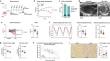
Anthracyclines are highly potent anti-cancer drugs, but their clinical use is limited by severe cardiotoxic side effects. The impact of anthracycline-induced cardiotoxicity (AIC) on left ventricular (LV) microarchitecture and diffusion properties remains unknown. This study sought to characterize AIC by cardiovascular magnetic resonance diffusion tensor imaging (DTI). Mice were treated with Doxorubicin (DOX; n = 16) for induction of AIC or saline as corresponding control (n = 15). Cardiac function was assessed via echocardiography at the end of the study period. Whole hearts (n = 8 per group) were scanned ex vivo by high-resolution DTI at 7 T. Results were correlated with histopathology and mass spectrometry imaging. Mice with AIC demonstrated systolic dysfunction (LVEF 52 ± 3% vs. 43 ± 6%, P < 0.001), impaired global longitudinal strain (−19.6 ± 2.0% vs. −16.6 ± 3.0%, P < 0.01), and cardiac atrophy (LV mass index [mg/mm], 4.3 ± 0.1 vs. 3.6 ± 0.2, P < 0.01). Regional sheetlet angles were significantly lower in AIC, whereas helix angle and relative helicity remained unchanged. In AIC, fractional anisotropy was increased (0.12 ± 0.01 vs. 0.14 ± 0.02, P < 0.05). DOX-treated mice displayed higher planar and less spherical anisotropy (CPlanar 0.07 ± 0.01 vs. 0.09 ± 0.01, P < 0.01; CSpherical 0.89 ± 0.01 vs. 0.87 ± 0.02, P < 0.05). CPlanar and CSpherical yielded good discriminatory power to distinguish between mice with and without AIC (c-index 0.91 and 0.84, respectively, P for both < 0.05). AIC is associated with regional changes in sheetlet angle but no major abnormalities of global LV microarchitecture. The geometric shape of the diffusion tensor is altered in AIC. DTI may provide a new tool for myocardial characterization in patients with AIC, which warrants future clinical studies to evaluate its diagnostic utility.
中文翻译:
通过弥散张量磁共振成像表征蒽环类药物诱导的心脏毒性
蒽环类药物是高效的抗癌药物,但其临床应用受到严重心脏毒性副作用的限制。蒽环类药物诱导的心脏毒性 (AIC) 对左心室 (LV) 微结构和扩散特性的影响仍然未知。本研究试图通过心血管磁共振弥散张量成像 (DTI) 来表征 AIC。小鼠用阿霉素 (DOX;n = 16) 用于诱导 AIC 或生理盐水作为相应的对照 (n = 15)。在研究期结束时通过超声心动图评估心脏功能。全心脏 (每组 n = 8) 在 7 T 处通过高分辨率 DTI 离体扫描,结果与组织病理学和质谱成像相关。AIC 小鼠表现出收缩功能障碍 (LVEF 52 ± 3% vs. 43 ± 6%,P < 0.001),整体纵向应变受损 (-19.6 ± 2.0% vs. -16.6 ± 3.0%,P < 0.01)和心脏萎缩 (LV 质量指数 [mg/mm],4.3 ± 0.1 vs. 3.6 ± 0.2,P < 0.01)。 在 AIC 中,区域片张角显著较低,而螺旋角和相对螺旋度保持不变。在 AIC 中,分数各向异性增加 (0.12 ± 0.01 vs. 0.14 ± 0.02,P < 0.05)。 DOX 处理的小鼠表现出较高的平面各向异性和较低的球面各向异性 (C平面 0.07 ± 0.01 vs. 0.09 ± 0.01,P < 0.01; C球形 0.89 ± 0.01 vs. 0.87 ± 0.02,P < 0.05)。 CPlanar 和 CSpherical 具有良好的区分能力,可以区分有和没有 AIC 的小鼠 (c 指数分别为 0.91 和 0.84,< 的 P 均为 0.05)。 AIC 与小片角的局部变化相关,但整体 LV 微结构无重大异常。扩散张量的几何形状在 AIC 中发生了变化。DTI 可能为 AIC 患者的心肌特征描述提供一种新工具,这需要未来的临床研究来评估其诊断效用。


 京公网安备 11010802027423号