Surgical and Radiologic Anatomy ( IF 1.2 ) Pub Date : 2024-02-20 , DOI: 10.1007/s00276-024-03303-2
Akino Aoki 1 , Yukiko Makihara 1 , Akihiro Tamura 1 , Takaya Ishii 2 , Kyutaro Kawagishi 2

|
Purpose
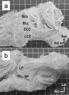
The calcaneocuboid joint is located in the lateral part of the foot and acts as a major stabilizer for the foot. Injuries to this joint often occur in association with ankle or foot injuries and are frequently overlooked, subsequently causing chronic pain or osteoarthritis. However, the relationship between ligaments surrounding the joint and joint instability remains unclear. Therefore, this study aimed to clarify the morphology and position of the ligaments surrounding the calcaneocuboid joint, and to reveal the relationship between the ligament structure.
Methods
The position and morphology of the bifurcate ligament (subdivided into calcaneonavicular and calcaneocuboid ligaments), dorsal calcaneocuboid ligament, lateral calcaneocuboid ligament, long plantar ligament, and short plantar ligament were measured (N = 11 feet in 6 Japanese cadavers). The circumference of the joint was quartered, while the ligament-uncovered area and the estimated cross-sectional area of each ligament were compared between the four sides. Furthermore, the estimated cross-sectional area of each ligament was calculated as an index for the ligament strength.
Results
The inferolateral side of the calcaneocuboid joint had the most uncovered area (54.63%) by the ligaments. In addition, the cross-sectional area of the ligaments on the lateral side was considerably smaller than that on the medial side.
Conclusion
Our results suggest that ligament weakness on the inferolateral side may cause instability of the calcaneocuboid joint, especially after an inversion sprain injury, and may decrease the lateral longitudinal arch function, which results in chronic foot pain.
中文翻译:

跟骰关节周围韧带的解剖分析对足部稳定性作用的影响
目的
跟骰关节位于脚的外侧部分,充当脚的主要稳定器。该关节的损伤通常与脚踝或足部损伤有关,并且经常被忽视,从而导致慢性疼痛或骨关节炎。然而,关节周围韧带与关节不稳定之间的关系仍不清楚。因此,本研究旨在明确跟骰关节周围韧带的形态和位置,揭示韧带结构之间的关系。
方法
测量了分叉韧带(分为跟舟韧带和跟骰韧带)、跟骰背韧带、跟骰外侧韧带、足底长韧带和足底短韧带的位置和形态(N = 6 具日本尸体中的 11 英尺)。将关节的周长四等分,同时比较四侧之间的韧带未覆盖面积和每个韧带的估计横截面积。此外,计算各韧带的估计横截面积作为韧带强度的指标。
结果
跟骰关节的下外侧被韧带覆盖的面积最多(54.63%)。此外,外侧韧带的横截面积明显小于内侧韧带的横截面积。
结论
我们的研究结果表明,下外侧韧带无力可能会导致跟骰关节不稳定,尤其是在内翻扭伤损伤后,并且可能会降低外侧纵弓功能,从而导致慢性足部疼痛。


 京公网安备 11010802027423号