当前位置:
X-MOL 学术
›
Nat. Commun.
›
论文详情
Our official English website, www.x-mol.net, welcomes your
feedback! (Note: you will need to create a separate account there.)
Graphene-enabled electron microscopy and correlated super-resolution microscopy of wet cells.
Nature Communications ( IF 14.7 ) Pub Date : 2015-Jun-11 , DOI: 10.1038/ncomms8384 Michal Wojcik , Margaret Hauser , Wan Li , Seonah Moon , Ke Xu
Nature Communications ( IF 14.7 ) Pub Date : 2015-Jun-11 , DOI: 10.1038/ncomms8384 Michal Wojcik , Margaret Hauser , Wan Li , Seonah Moon , Ke Xu

|
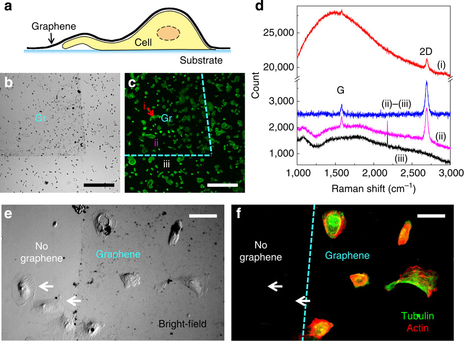
The application of electron microscopy to hydrated biological samples has been limited by high-vacuum operating conditions. Traditional methods utilize harsh and laborious sample dehydration procedures, often leading to structural artefacts and creating difficulties for correlating results with high-resolution fluorescence microscopy. Here, we utilize graphene, a single-atom-thick carbon meshwork, as the thinnest possible impermeable and conductive membrane to protect animal cells from vacuum, thus enabling high-resolution electron microscopy of wet and untreated whole cells with exceptional ease. Our approach further allows for facile correlative super-resolution and electron microscopy of wet cells directly on the culturing substrate. In particular, individual cytoskeletal actin filaments are resolved in hydrated samples through electron microscopy and well correlated with super-resolution results.
中文翻译:

启用石墨烯的电子显微镜和湿细胞的相关超分辨率显微镜。
电子显微镜在水合生物样品中的应用受到高真空操作条件的限制。传统方法利用苛刻且费力的样品脱水程序,这通常会导致结构伪影,并给将结果与高分辨率荧光显微镜相关联带来困难。在这里,我们利用石墨烯(一种单原子厚度的碳网)作为最薄的不可渗透和导电的膜来保护动物细胞免受真空侵害,从而使湿的和未处理的整个细胞的高分辨率电子显微镜检查变得异常轻松。我们的方法进一步允许直接在培养基质上对湿细胞进行简便的相关超分辨率和电子显微镜检查。尤其是,
更新日期:2015-06-15
中文翻译:

启用石墨烯的电子显微镜和湿细胞的相关超分辨率显微镜。
电子显微镜在水合生物样品中的应用受到高真空操作条件的限制。传统方法利用苛刻且费力的样品脱水程序,这通常会导致结构伪影,并给将结果与高分辨率荧光显微镜相关联带来困难。在这里,我们利用石墨烯(一种单原子厚度的碳网)作为最薄的不可渗透和导电的膜来保护动物细胞免受真空侵害,从而使湿的和未处理的整个细胞的高分辨率电子显微镜检查变得异常轻松。我们的方法进一步允许直接在培养基质上对湿细胞进行简便的相关超分辨率和电子显微镜检查。尤其是,


 京公网安备 11010802027423号