CardioVascular and Interventional Radiology ( IF 2.8 ) Pub Date : 2024-01-23 , DOI: 10.1007/s00270-023-03648-y
Geoffrey Bourgeais 1 , Eric Frampas 1 , Renan Liberge 1 , Aymeric Nicolas 1 , Claire Defrance 1 , François-Xavier Blanc 2 , Sandrine Coudol 3 , Olivier Morla 1

|
Purpose
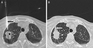
To determine whether instillation of normal saline solution for sealing the needle track reduces incidence of pneumothorax and chest tube placement after computed tomography-guided percutaneous lung biopsy.
Materials and Methods
A total of 242 computed tomography-guided percutaneous lung biopsies performed at a single institution were retrospectively reviewed, including 93 biopsies in which the needle track was sealed by instillation of 3–5 ml of normal saline solution during needle withdrawal (water seal group) and 149 biopsies without sealing (control group). Patient and lesion characteristics, procedure-specific variables, pneumothorax and chest tube placement rates were recorded.
Results
Baseline characteristics were comparable in both groups. There was a statistically significant decrease in the pneumothorax rate (19.4% [18/93] vs. 40.9% [61/149]; p < 0.001) and a numerically lower chest tube placement rate without significant reduction (4.3% [4/93] vs. 10.7% [16/149]; p = 0.126) with using normal saline instillation for sealing the needle track versus not using sealant material. Using a multiple logistic regression analysis, using normal saline instillation to seal the needle track, having a senior radiologist as operator of the procedure and putting patients in prone position were significantly associated with a decreased risk of pneumothorax. The presence of emphysema along the needle track was significantly associated with an increased risk of pneumothorax. No complication was observed due to normal saline injection.
Conclusion
Normal saline solution instillation for sealing the needle track after computed tomography-guided percutaneous lung biopsy is a simple, low-cost and safe technique resulted in significantly decreased pneumothorax occurrence and a numerically lower chest tube placement rate, and might help to reduce both hospitalization risks and costs for the healthcare system.
Level of evidence 3 Non-controlled retrospective cohort study.
Graphical Abstract
中文翻译:

计算机断层扫描引导下经皮肺活检后滴注生理盐水封闭针道的气胸发生率
目的
确定在计算机断层扫描引导的经皮肺活检后,滴注生理盐水溶液以密封针道是否可以减少气胸和胸管放置的发生率。
材料和方法
回顾性分析同一机构进行的242例CT引导下经皮肺活检,其中93例为退针时滴注3~5 ml生理盐水封闭针道的活检(水封组), 149 个未密封的活检组织(对照组)。记录患者和病变特征、手术特定变量、气胸和胸管放置率。
结果
两组的基线特征相当。气胸率显着下降(19.4% [18/93] vs. 40.9% [61/149]; p < 0.001),胸管置入率在数字上较低,但没有显着下降(4.3% [4/93]) ] vs. 10.7% [16/149]; p = 0.126) 使用生理盐水滴注密封针道与不使用密封剂材料相比。使用多重逻辑回归分析,使用生理盐水滴注来密封针道,由高级放射科医生作为手术操作员以及让患者处于俯卧位与气胸风险降低显着相关。沿针道出现肺气肿与气胸风险增加显着相关。注射生理盐水未观察到并发症。
结论
计算机断层扫描引导下经皮肺活检后滴注生理盐水密封针道是一种简单、低成本且安全的技术,可显着减少气胸发生率和较低的胸管放置率,并可能有助于降低住院风险以及医疗保健系统的成本。
证据级别 3非对照回顾性队列研究。


 京公网安备 11010802027423号