Our official English website, www.x-mol.net, welcomes your
feedback! (Note: you will need to create a separate account there.)
An Electroencephalography Profile of Paroxysmal Kinesigenic Dyskinesia
Advanced Science ( IF 14.3 ) Pub Date : 2024-01-16 , DOI: 10.1002/advs.202306321 Huichun Luo 1, 2 , Xiaojun Huang 1 , Ziyi Li 1 , Wotu Tian 1 , Kan Fang 1, 3 , Taotao Liu 1 , Shige Wang 1 , Beisha Tang 4 , Ji Hu 5 , Ti-Fei Yuan 2, 6, 7 , Li Cao 1, 8
Advanced Science ( IF 14.3 ) Pub Date : 2024-01-16 , DOI: 10.1002/advs.202306321 Huichun Luo 1, 2 , Xiaojun Huang 1 , Ziyi Li 1 , Wotu Tian 1 , Kan Fang 1, 3 , Taotao Liu 1 , Shige Wang 1 , Beisha Tang 4 , Ji Hu 5 , Ti-Fei Yuan 2, 6, 7 , Li Cao 1, 8
Affiliation

|
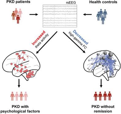
Paroxysmal kinesigenic dyskinesia (PKD) is associated with a disturbance of neural circuit and network activities, while its neurophysiological characteristics have not been fully elucidated. This study utilized the high-density electroencephalogram (hd-EEG) signals to detect abnormal brain activity of PKD and provide a neural biomarker for its clinical diagnosis and PKD progression monitoring. The resting hd-EEGs are recorded from two independent datasets and then source-localized for measuring the oscillatory activities and function connectivity (FC) patterns of cortical and subcortical regions. The abnormal elevation of theta oscillation in wildly brain regions represents the most remarkable physiological feature for PKD and these changes returned to healthy control level in remission patients. Another remarkable feature of PKD is the decreased high-gamma FCs in non-remission patients. Subtype analyses report that increased theta oscillations may be related to the emotional factors of PKD, while the decreased high-gamma FCs are related to the motor symptoms. Finally, the authors established connectome-based predictive modelling and successfully identified the remission state in PKD patients in dataset 1 and dataset 2. The findings establish a clinically relevant electroencephalography profile of PKD and indicate that hd-EEG can provide robust neural biomarkers to evaluate the prognosis of PKD.
中文翻译:

阵发性运动机能障碍的脑电图概况
阵发性运动动力学障碍 (PKD) 与神经回路和网络活动紊乱有关,而其神经生理学特征尚未完全阐明。本研究利用高密度脑电图 (hd-EEG) 信号检测 PKD 的异常脑活动,为其临床诊断和 PKD 进展监测提供神经生物标志物。静息 hd-EEG 从两个独立的数据集中记录下来,然后进行源定位,用于测量皮层和皮层下区域的振荡活动和功能连接 (FC) 模式。狂野脑区 θ 振荡的异常升高是 PKD 最显著的生理特征,这些变化在缓解期患者中恢复到健康的控制水平。PKD 的另一个显着特征是非缓解患者的高 γ FC 减少。亚型分析报告称,θ 振荡增加可能与 PKD 的情绪因素有关,而高 γ FC 减少与运动症状有关。最后,作者建立了基于连接组的预测模型,并在数据集 1 和数据集 2 中成功确定了 PKD 患者的缓解状态。研究结果建立了 PKD 的临床相关脑电图概况,并表明 hd-EEG 可以提供强大的神经生物标志物来评估 PKD 的预后。
更新日期:2024-01-16
中文翻译:

阵发性运动机能障碍的脑电图概况
阵发性运动动力学障碍 (PKD) 与神经回路和网络活动紊乱有关,而其神经生理学特征尚未完全阐明。本研究利用高密度脑电图 (hd-EEG) 信号检测 PKD 的异常脑活动,为其临床诊断和 PKD 进展监测提供神经生物标志物。静息 hd-EEG 从两个独立的数据集中记录下来,然后进行源定位,用于测量皮层和皮层下区域的振荡活动和功能连接 (FC) 模式。狂野脑区 θ 振荡的异常升高是 PKD 最显著的生理特征,这些变化在缓解期患者中恢复到健康的控制水平。PKD 的另一个显着特征是非缓解患者的高 γ FC 减少。亚型分析报告称,θ 振荡增加可能与 PKD 的情绪因素有关,而高 γ FC 减少与运动症状有关。最后,作者建立了基于连接组的预测模型,并在数据集 1 和数据集 2 中成功确定了 PKD 患者的缓解状态。研究结果建立了 PKD 的临床相关脑电图概况,并表明 hd-EEG 可以提供强大的神经生物标志物来评估 PKD 的预后。


 京公网安备 11010802027423号