Journal of Digital Imaging ( IF 2.9 ) Pub Date : 2024-01-10 , DOI: 10.1007/s10278-023-00929-3
Chih-Hung Wang, Jia-Da Li, Cheng-Yi Wu, Yu-Chen Wu, Joyce Tay, Meng-Che Wu, Ching-Hang Hsu, Yi-Kuan Liu, Chu-Song Chen, Chien-Hua Huang
|
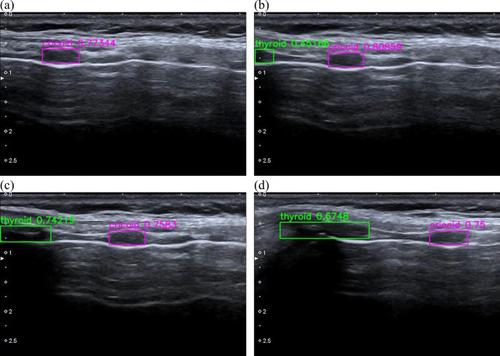
We aimed to develop machine learning (ML)-based algorithms to assist physicians in ultrasound-guided localization of cricoid cartilage (CC) and thyroid cartilage (TC) in cricothyroidotomy. Adult female volunteers were prospectively recruited from two hospitals between September and December, 2020. Ultrasonographic images were collected via a modified longitudinal technique. You Only Look Once (YOLOv5s), Faster Regions with Convolutional Neural Network features (Faster R-CNN), and Single Shot Detector (SSD) were selected as the model architectures. A total of 488 women (mean age: 36.0 years) participated in the study, contributing to a total of 292,053 frames of ultrasonographic images. The derived ML-based algorithms demonstrated excellent discriminative performance for the presence of CC (area under the receiver operating characteristic curve [AUC]: YOLOv5s, 0.989, 95% confidence interval [CI]: 0.982–0.994; Faster R-CNN, 0.986, 95% CI: 0.980–0.991; SSD, 0.968, 95% CI: 0.956–0.977) and TC (AUC: YOLOv5s, 0.989, 95% CI: 0.977–0.997; Faster R-CNN, 0.981, 95% CI: 0.965–0.991; SSD, 0.982, 95% CI: 0.973–0.990). Furthermore, in the frames where the model could correctly indicate the presence of CC or TC, it also accurately localized CC (intersection-over-union: YOLOv5s, 0.753, 95% CI: 0.739–0.765; Faster R-CNN, 0.720, 95% CI: 0.709–0.732; SSD, 0.739, 95% CI: 0.726–0.751) or TC (intersection-over-union: YOLOv5s, 0.739, 95% CI: 0.722–0.755; Faster R-CNN, 0.709, 95% CI: 0.687–0.730; SSD, 0.713, 95% CI: 0.695–0.730). The ML-based algorithms could identify anatomical landmarks for cricothyroidotomy in adult females with favorable discriminative and localization performance. Further studies are warranted to transfer this algorithm to hand-held portable ultrasound devices for clinical use.
中文翻译:
机器学习在超声检查中的应用,用于识别女性成人环甲膜切开术的解剖标志:一项多中心前瞻性观察研究
我们的目标是开发基于机器学习 (ML) 的算法,以协助医生在环甲膜切开术中超声引导下定位环状软骨 (CC) 和甲状软骨 (TC)。 2020 年 9 月至 12 月期间,前瞻性地从两家医院招募成年女性志愿者。通过改进的纵向技术收集超声图像。选择 You Only Look Once (YOLOv5s)、Faster Regions with Convolutional Neural Network features (Faster R-CNN) 和 Single Shot Detector (SSD) 作为模型架构。共有 488 名女性(平均年龄:36.0 岁)参与了这项研究,共贡献了 292,053 帧超声图像。派生的基于 ML 的算法对 CC 的存在表现出出色的判别性能(受试者工作特征曲线下面积 [AUC]:YOLOv5s,0.989,95% 置信区间 [CI]:0.982–0.994;Faster R-CNN,0.986, 95% CI:0.980–0.991;SSD,0.968,95% CI:0.956–0.977)和 TC(AUC:YOLOv5s,0.989,95% CI:0.977–0.997;Faster R-CNN,0.981,95% CI:0.965– 0.991;SSD,0.982,95% CI:0.973–0.990)。此外,在模型能够正确指示 CC 或 TC 存在的帧中,它还准确地定位了 CC(交集交集:YOLOv5s,0.753,95% CI:0.739–0.765;Faster R-CNN,0.720,95) % CI:0.709–0.732;SSD,0.739,95% CI:0.726–0.751)或 TC(交集:YOLOv5s,0.739,95% CI:0.722–0.755;Faster R-CNN,0.709,95% CI :0.687–0.730;SSD,0.713,95% CI:0.695–0.730)。基于机器学习的算法可以识别成年女性环甲膜切开术的解剖标志,具有良好的判别和定位性能。 需要进一步研究将该算法转移到手持式便携式超声设备以供临床使用。


 京公网安备 11010802027423号