Pediatric Radiology ( IF 2.1 ) Pub Date : 2024-01-09 , DOI: 10.1007/s00247-023-05848-7
Helio V Neves da Silva 1, 2, 3 , Jason P Weinman 1, 2 , Erin K Englund 1 , Robin R Deterding 2, 4 , Dunbar D Ivy 2, 5 , Lorna P Browne 1, 2
|
Background
Mutations in the T-Box 4 (TBX4) gene are a lesser-known cause of heritable pulmonary arterial hypertension (PAH). Patients with heritable PAH typically have worse outcomes when compared with patients with idiopathic PAH, yet little is known about the phenotypical presentation of this mutation.
Objective
This article reviews the pattern of chest CT findings in pediatric patients with PAH and TBX4 mutations and compares their radiographic presentation with those of age-matched patients with PAH but without TBX4 mutations.
Materials and methods
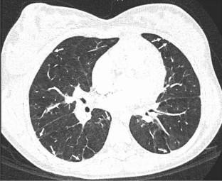
A retrospective chart review of the pulmonary arterial hypertension database was performed. Pediatric patients with PAH-confirmed TBX4 mutations and an available high CT were included. Fifteen (9 females) patients met the inclusion criteria. Fourteen (8 females) age-matched controls with diagnosed PAH but without TBX4 mutations were also evaluated. The median age at diagnosis was 7.4 years (range: 0.1–16.4 years). Demographic information and clinical outcomes were collected. CTs of the chest were reviewed for multiple airway, parenchymal, and structural abnormalities (16 imaging findings in total). Chi-square tests were used to compare the prevalence of each imaging finding in the TBX4 cohort compared to the control group.
Results
Patients with TBX-4 mutations had increased presence of peripheral or subpleural irregularity (73% vs 0%, P < 0.01), cystic lucencies (67% vs 7%, P < 0.01), and linear or reticular opacity (53% vs 0%, P < 0.01) compared to the control group. Ground glass opacities, bronchiectasis, and centrilobular nodules were not significantly different between the two patient groups (P > 0.05).
Conclusion
TBX4 mutations have distinct imaging phenotypes in pediatric patients with PAH. Compared to patients without this mutation, patients with TBX-4 genes typically present with peripheral or subpleural irregularity, cystic lucencies, and linear or reticular opacity.
Graphical Abstract
中文翻译:
TBX4 突变的计算机断层扫描结果:儿童严重肺动脉高压的常见原因
背景
T-Box 4 (TBX4) 基因突变是遗传性肺动脉高压 (PAH) 的一个鲜为人知的原因。与特发性 PAH 患者相比,遗传性 PAH 患者的预后通常较差,但人们对这种突变的表型表现知之甚少。
客观的
本文回顾了患有 PAH 和 TBX4 突变的儿科患者的胸部 CT 表现,并将其影像学表现与年龄匹配的 PAH 但无 TBX4 突变的患者的影像学表现进行了比较。
材料和方法
对肺动脉高压数据库进行了回顾性图表审查。患有 PAH 确诊的 TBX4 突变且具有可用高 CT 的儿科患者也被纳入其中。十五名(9 名女性)患者符合纳入标准。还评估了 14 名(8 名女性)年龄匹配的被诊断为 PAH 但没有 TBX4 突变的对照。诊断时的中位年龄为 7.4 岁(范围:0.1-16.4 岁)。收集人口统计信息和临床结果。检查胸部 CT 是否有多个气道、实质和结构异常(总共 16 项影像学发现)。卡方检验用于比较 TBX4 队列与对照组中每种影像学发现的发生率。
结果
TBX-4突变患者的外周或胸膜下不规则性(73% vs 0%, P < 0.01)、囊性透明(67% vs 7%, P < 0.01)以及线状或网状混浊(53% vs 0)的发生率增加%, P < 0.01)与对照组相比。两组患者毛玻璃影、支气管扩张和小叶中心结节差异无统计学意义( P > 0.05)。
结论
TBX4 突变在儿科 PAH 患者中具有独特的影像表型。与没有这种突变的患者相比,具有 TBX-4 基因的患者通常表现为外周或胸膜下不规则、囊性透明以及线性或网状混浊。


 京公网安备 11010802027423号