当前位置:
X-MOL 学术
›
Dev. Biol.
›
论文详情
Our official English website, www.x-mol.net, welcomes your
feedback! (Note: you will need to create a separate account there.)
Claudin-3 in the non-neural ectoderm is essential for neural fold fusion in chicken embryos
Developmental Biology ( IF 2.5 ) Pub Date : 2023-12-27 , DOI: 10.1016/j.ydbio.2023.12.009 Elizabeth-Ann Legere 1 , Amanda I Baumholtz 1 , Jean-François Boisclair Lachance 2 , Madison Archer 3 , Jörg Piontek 4 , Aimee K Ryan 1
Developmental Biology ( IF 2.5 ) Pub Date : 2023-12-27 , DOI: 10.1016/j.ydbio.2023.12.009 Elizabeth-Ann Legere 1 , Amanda I Baumholtz 1 , Jean-François Boisclair Lachance 2 , Madison Archer 3 , Jörg Piontek 4 , Aimee K Ryan 1
Affiliation
|
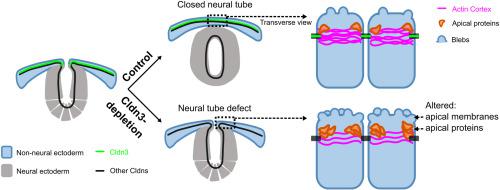
The neural tube, the embryonic precursor to the brain and spinal cord, begins as a flat sheet of epithelial cells, divided into non-neural and neural ectoderm. Proper neural tube closure requires that the edges of the neural ectoderm, the neural folds, to elevate upwards and fuse along the dorsal midline of the embryo. We have previously shown that members of the claudin protein family are required for the early phases of chick neural tube closure. Claudins are transmembrane proteins, localized in apical tight junctions within epithelial cells where they are essential for regulation of paracellular permeability, strongly involved in apical-basal polarity, cell-cell adhesion, and bridging the tight junction to cytoplasmic proteins. Here we explored the role of Claudin-3 (Cldn3), which is specifically expressed in the non-neural ectoderm. We discovered that depletion of Cldn3 causes folic acid-insensitive primarily spinal neural tube defects due to a failure in neural fold fusion. Apical cell surface morphology of Cldn3-depleted non-neural ectodermal cells exhibited increased membrane blebbing and smaller apical surfaces. Although apical-basal polarity was retained, we observed altered Par3 and Pals1 protein localization patterns within the apical domain of the non-neural ectodermal cells in Cldn3-depleted embryos. Furthermore, F-actin signal was reduced at apical junctions. Our data presents a model of spina bifida, and the role that Cldn3 is playing in regulating essential apical cell processes in the non-neural ectoderm required for neural fold fusion.
中文翻译:

非神经外胚层中的 Claudin-3 对于鸡胚胎中的神经褶皱融合至关重要
神经管是大脑和脊髓的胚胎前体,最初是一块扁平的上皮细胞,分为非神经和神经外胚层。适当的神经管闭合要求神经外胚层的边缘(神经褶皱)向上抬起并沿着胚胎的背中线融合。我们之前已经表明,小鸡神经管闭合的早期阶段需要 claudin 蛋白家族的成员。Claudins 是跨膜蛋白,位于上皮细胞内的顶端紧密连接处,它们对调节细胞旁通透性至关重要,强烈参与顶端-基底极性、细胞间粘附,并桥接紧密连接与细胞质蛋白。在这里,我们探讨了 Claudin-3 (Cldn3) 的作用,它在非神经外胚层中特异性表达。我们发现 Cldn3 的耗竭由于神经皱襞融合失败导致叶酸不敏感,主要是脊髓神经管缺陷。Cldn3 耗尽的非神经外胚层细胞的顶端细胞表面形态表现出膜起泡增加和顶端表面较小。尽管保留了顶端-基底极性,但我们观察到在 Cldn3 耗尽的胚胎中,非神经外胚层细胞顶端结构域内的 Par3 和 Pals1 蛋白定位模式发生了变化。此外,F-肌动蛋白信号在顶端连接处减弱。我们的数据提出了脊柱裂模型,以及 Cldn3 在调节神经折叠融合所需的非神经外胚层中的基本顶细胞过程中的作用。
更新日期:2023-12-27
中文翻译:
非神经外胚层中的 Claudin-3 对于鸡胚胎中的神经褶皱融合至关重要
神经管是大脑和脊髓的胚胎前体,最初是一块扁平的上皮细胞,分为非神经和神经外胚层。适当的神经管闭合要求神经外胚层的边缘(神经褶皱)向上抬起并沿着胚胎的背中线融合。我们之前已经表明,小鸡神经管闭合的早期阶段需要 claudin 蛋白家族的成员。Claudins 是跨膜蛋白,位于上皮细胞内的顶端紧密连接处,它们对调节细胞旁通透性至关重要,强烈参与顶端-基底极性、细胞间粘附,并桥接紧密连接与细胞质蛋白。在这里,我们探讨了 Claudin-3 (Cldn3) 的作用,它在非神经外胚层中特异性表达。我们发现 Cldn3 的耗竭由于神经皱襞融合失败导致叶酸不敏感,主要是脊髓神经管缺陷。Cldn3 耗尽的非神经外胚层细胞的顶端细胞表面形态表现出膜起泡增加和顶端表面较小。尽管保留了顶端-基底极性,但我们观察到在 Cldn3 耗尽的胚胎中,非神经外胚层细胞顶端结构域内的 Par3 和 Pals1 蛋白定位模式发生了变化。此外,F-肌动蛋白信号在顶端连接处减弱。我们的数据提出了脊柱裂模型,以及 Cldn3 在调节神经折叠融合所需的非神经外胚层中的基本顶细胞过程中的作用。


 京公网安备 11010802027423号