Basic Research in Cardiology ( IF 7.5 ) Pub Date : 2023-12-26 , DOI: 10.1007/s00395-023-01029-7 Gu Li , He Huang , Yanshuang Wu , Chang Shu , Narae Hwang , Qifei Li , Rose Zhao , Hilaire C. Lam , William M. Oldham , Souheil EI-Chemaly , Pankaj B. Agrawal , Jie Tian , Xiaoli Liu , Mark A. Perrella
|
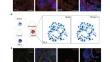
A deficiency of striated preferentially expressed gene (Speg), a member of the myosin light chain kinase family, results in abnormal myofibril structure and function of immature cardiomyocytes (CMs), corresponding with a dilated cardiomyopathy, heart failure and perinatal death. Mitochondrial development plays a role in cardiomyocyte maturation. Therefore, this study investigated whether Speg deficiency ( – / – ) in CMs would result in mitochondrial abnormalities. Speg wild-type and Speg−/− C57BL/6 littermate mice were utilized for assessment of mitochondrial structure by transmission electron and confocal microscopies. Speg was expressed in the first and second heart fields at embryonic (E) day 7.5, prior to the expression of mitochondrial Na+/Ca2+/Li+ exchanger (NCLX) at E8.5. Decreases in NCLX expression (E11.5) and the mitochondrial-to-nuclear DNA ratio (E13.5) were observed in Speg−/− hearts. Imaging of E18.5 Speg−/− hearts revealed abnormal mitochondrial cristae, corresponding with decreased ATP production in cells fed glucose or palmitate, increased levels of mitochondrial superoxide and depolarization of mitochondrial membrane potential. Interestingly, phosphorylated (p) PGC-1α, a key mediator of mitochondrial development, was significantly reduced in Speg−/− hearts during screening for targeted genes. Besides Z-line expression, Speg partially co-localized with PGC-1α in the sarcomeric region and was found in the same complex by co-immunoprecipitation. Overexpression of a Speg internal serine/threonine kinase domain in Speg−/− CMs promoted translocation of pPGC-1α into the nucleus, and restored ATP production that was abolished by siRNA-mediated silencing of PGC-1α. Our results demonstrate a critical role of Speg in mitochondrial development and energy metabolism in CMs, mediated in part by phosphorylation of PGC-1α.
中文翻译:
横纹优先表达基因缺陷导致发育中的心肌细胞线粒体功能障碍
横纹优先表达基因 ( Speg )(肌球蛋白轻链激酶家族的成员)的缺陷会导致未成熟心肌细胞 (CM) 的肌原纤维结构和功能异常,从而导致扩张型心肌病、心力衰竭和围产期死亡。线粒体发育在心肌细胞成熟中发挥作用。因此,本研究调查了CM 中Speg缺乏 (-/-) 是否会导致线粒体异常。Speg野生型和Speg −/− C57BL/6 同窝小鼠用于通过透射电子和共聚焦显微镜评估线粒体结构。 Speg 在胚胎 ( E ) 第 7.5 天在第一和第二心田中表达,然后在 E8.5 表达线粒体 Na + /Ca2 + /Li +交换器 (NCLX)。在Speg −/−心脏中观察到 NCLX 表达 (E11.5) 和线粒体与核 DNA 比率 (E13.5) 的降低。 E18.5 Speg -/−心脏的成像显示异常的线粒体嵴,与喂食葡萄糖或棕榈酸盐的细胞中 ATP 产生减少、线粒体超氧化物水平增加和线粒体膜电位去极化相对应。有趣的是,在筛选目标基因期间,Speg −/−心脏中磷酸化 ( p ) PGC-1α(线粒体发育的关键介质)显着减少。除了 Z 线表达外,Speg 与 PGC-1α 部分共定位于肌节区域,并通过免疫共沉淀发现在同一复合物中。Speg −/− CM中 Speg 内部丝氨酸/苏氨酸激酶结构域的过度表达促进了 pPGC-1α 易位到细胞核中,并恢复了因 siRNA 介导的 PGC-1α 沉默而消除的 ATP 产生。我们的结果证明 Speg 在 CM 线粒体发育和能量代谢中发挥关键作用,部分由 PGC-1α 磷酸化介导。


 京公网安备 11010802027423号