Scientific Reports ( IF 3.8 ) Pub Date : 2023-12-08 , DOI: 10.1038/s41598-023-48776-0
Jon W S McCullough 1 , Peter V Coveney 1, 2, 3
|
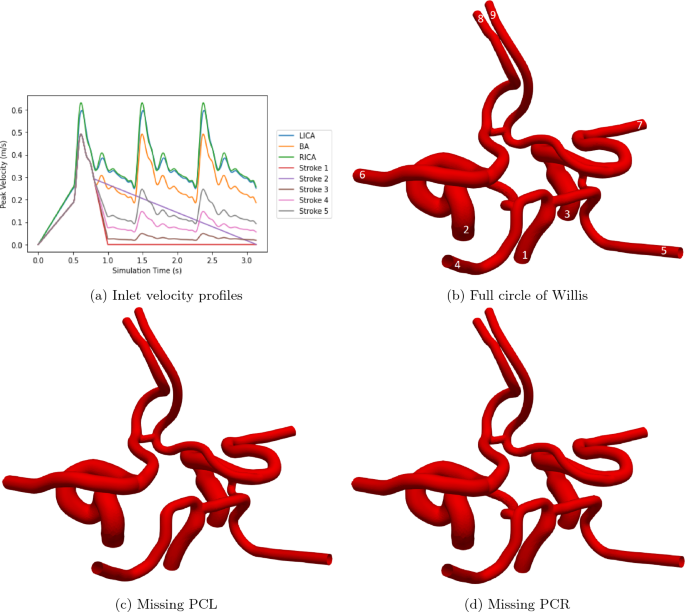
On a global scale, cerebro- and cardiovascular diseases have long been one of the leading causes of death and disability and their prevalence appears to be increasing in recent times. Understanding potential biomarkers and risk factors will help to identify individuals potentially at risk of suffering an ischemic stroke. However, the widely variable construction of the cerebral vasculature makes it difficult to provide a specific assessment without the knowledge of a patient’s physiology. In this paper we use the 3D blood flow simulator HemeLB to study flow within three common structural variations of the circle of Willis during and in the moments after a blockage of the basilar artery. This tool, based on the lattice Boltzmann method, allows the 3D flow entering the basilar artery to be finely controlled to replicate the cessation of blood feeding this particular vessel—we demonstrate this with several examples including a sudden halt to flow and a gradual loss of flow over three heartbeat cycles. In this work we start with an individualised 3D representation of a full circle of Willis and then construct two further domains by removing the left or right posterior communicating arteries from this geometry. Our results indicate how, and how quickly, the circle of Willis is able to redistribute flow following such a stroke. Due to the choice of infarct, the greatest reduction in flow was observed in the posterior cerebral arteries where flow was reduced by up to 70% in some cases. The high resolution domains used in this study permit the velocity magnitude and wall shear stress to be analysed at key points during and following the stroke. The model we present here indicates how personalised vessels are required to provide the best insight into stroke risk for a given individual.
中文翻译:
基底动脉梗塞和 Willis 环内血流的高分辨率模拟
在全球范围内,脑和心血管疾病长期以来一直是死亡和残疾的主要原因之一,并且近年来其患病率似乎在增加。了解潜在的生物标志物和危险因素将有助于识别有可能患缺血性中风风险的个体。然而,脑脉管系统结构的广泛变化使得在不了解患者生理学的情况下很难提供具体的评估。在本文中,我们使用 3D 血流模拟器 HemeLB 研究基底动脉阻塞期间和阻塞后瞬间 Willis 环的三种常见结构变化内的血流。该工具基于格子玻尔兹曼方法,可以精细控制进入基底动脉的 3D 血流,以复制该特定血管的血液供给停止情况 - 我们通过几个例子来证明这一点,包括血流突然停止和血流逐渐丧失流过三个心跳周期。在这项工作中,我们从 Willis 整圈的个性化 3D 表示开始,然后通过从该几何结构中移除左或右后交通动脉来构建另外两个域。我们的结果表明,威利斯环如何以及以多快的速度能够在此类中风后重新分配血流。由于梗塞的选择,在大脑后动脉中观察到流量减少最大,在某些情况下流量减少高达 70%。本研究中使用的高分辨率域允许在冲程期间和之后的关键点分析速度大小和壁剪切应力。 我们在此展示的模型表明如何需要个性化血管来提供对特定个体中风风险的最佳洞察。


 京公网安备 11010802027423号