Scientific Reports ( IF 3.8 ) Pub Date : 2023-11-27 , DOI: 10.1038/s41598-023-47619-2 Nutcha Yodrabum 1 , Irin Chaikangwan 1 , Jirapat Tianrungroj 1 , Songsak Suksantilap 2 , Suttichai Chalalaisathaphorn 2 , Palanan Siriwanarangsun 3

|
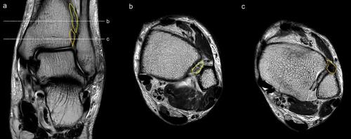
Preservation of syndesmotic ligaments is crucial for preventing adverse sequelae at the donor site following free fibula osteocutaneous flap harvesting. This study sought to determine the relationship between distal tibiofibular ligaments and the fibular segment to identify radiological landmarks that facilitate safe and precise flap. The distances between the distal tibiofibular ligaments (anterior inferior tibiofibular ligament [AITFL], posterior inferior tibiofibular ligament [PITFL]) and the fibular segment, as well as the lower border of the interosseous membrane, were measured on magnetic resonance imaging (MRI) scans of 296 patients without any perceivable ankle abnormalities. The mean distances (± SD) between the distal end of the fibula and the AITFL, PITFL, and lower interosseous membrane border were 3.0 ± 0.4 cm, 2.6 ± 0.4 cm, and 3.9 ± 0.6 cm, respectively. The distance between the talar dome and the PITFL exhibited a range of 0.0–0.5 cm. Our findings support preserving a distal fibular remnant of at least 4 cm to avoid injury to the syndesmotic ligament throughout fibula osteocutaneous flap harvesting. The talar dome could serve as a useful radiological landmark for identifying the upper border of PITFL during preoperative evaluation, and thus facilitating precise and safe flap procurement.
中文翻译:

磁共振成像联合韧带复合体的放射学标志与腓骨游离皮瓣采集程序相关
保留腓骨联合韧带对于预防游离腓骨骨皮瓣采集后供体部位出现不良后遗症至关重要。本研究试图确定远端下胫腓韧带和腓骨段之间的关系,以确定有助于安全和精确皮瓣移植的放射学标志。通过磁共振成像 (MRI) 扫描测量远端下胫腓韧带(前下胫腓韧带 [AITFL]、后下下胫腓韧带 [PITFL])与腓骨段以及骨间膜下缘之间的距离296 名没有任何明显踝关节异常的患者。腓骨远端与 AITFL、PITFL 和骨间膜下缘之间的平均距离 (± SD) 分别为 3.0 ± 0.4 cm、2.6 ± 0.4 cm 和 3.9 ± 0.6 cm。距骨穹顶和 PITFL 之间的距离范围为 0.0-0.5 cm。我们的研究结果支持保留至少 4 厘米的远端腓骨残余物,以避免在腓骨骨皮瓣采集过程中损伤韧带联合韧带。距骨穹顶可以作为术前评估期间识别 PITFL 上缘的有用放射学标志,从而促进精确、安全的皮瓣采购。


 京公网安备 11010802027423号