Pituitary ( IF 3.3 ) Pub Date : 2023-10-30 , DOI: 10.1007/s11102-023-01352-1
Meric Coskun 1 , Halit Nahit Sendur 2 , Mahi Nur Cerit 2 , Afruz Babayeva 1 , Ethem Turgay Cerit 1 , Mehmet Muhittin Yalcin 1 , Alev Eroglu Altinova 1 , Mujde Akturk 1 , Mehmet Ayhan Karakoc 1 , Fusun Balos Toruner 1
|
Purpose
The effects of acromegaly on soft tissues, bones and joints are well-documented, but information on its effects on muscle mass and quality remains limited. The primary goal of this study is to assess the sonoelastographic features of forearm muscles in patients with acromegaly.
Method
Forty-five patients with acromegaly and 45 healthy controls similar in terms of gender, age, and body mass index (BMI) were included in a single-center, multidisciplinary, cross-sectional study. The body composition was analyzed using bioelectrical impedance analysis (BIA), and height-adjusted appendicular skeletal muscle index (hSMI) was calculated. The dominant hand’s grip strength was also measured. Two radiologists specialized in the musculoskeletal system employed ultrasound shear wave elastography (SWE) to assess the thickness and stiffness of brachioradialis and biceps brachii muscles.
Results
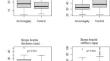
The acromegaly group had significantly higher thickness of both the biceps brachii (p = 0.034) and brachioradialis muscle (p = 0.046) than the control group. However, the stiffness of the biceps brachii (p = 0.001) and brachioradialis muscle (p = 0.001) was lower in the acromegaly group than in the control group. Disease activity has not caused a significant difference in muscle thickness and stiffness in the acromegaly group (p > 0.05). The acromegaly group had a higher hSMI (p = 0.004) than the control group. The hand grip strength was similar between the acromegaly and control group (p = 0.594).
Conclusion
The patients with acromegaly have an increased muscle thickness but decreased muscle stiffness in the forearm muscles responsible for elbow flexion. Acromegaly can lead to a permanent deterioration of the muscular structure regardless of the disease activity.
中文翻译:
超声剪切波弹性成像评估肢端肥大症患者前臂肌肉
目的
肢端肥大症对软组织、骨骼和关节的影响已有充分记录,但有关其对肌肉质量和质量影响的信息仍然有限。本研究的主要目标是评估肢端肥大症患者前臂肌肉的声弹性成像特征。
方法
一项单中心、多学科、横断面研究纳入了 45 名肢端肥大症患者和 45 名在性别、年龄和体重指数 (BMI) 方面相似的健康对照。使用生物电阻抗分析(BIA)分析身体成分,并计算高度调整的四肢骨骼肌指数(hSMI)。还测量了惯用手的握力。两名专门研究肌肉骨骼系统的放射科医生采用超声波剪切波弹性成像 (SWE) 来评估肱桡肌和肱二头肌的厚度和硬度。
结果
肢端肥大症组的肱二头肌 (p = 0.034) 和肱桡肌 (p = 0.046) 厚度均显着高于对照组。然而,肢端肥大症组的肱二头肌 (p = 0.001) 和肱桡肌 (p = 0.001) 的硬度低于对照组。疾病活动并未导致肢端肥大症组的肌肉厚度和硬度出现显着差异(p > 0.05)。肢端肥大症组的 hSMI (p = 0.004) 高于对照组。肢端肥大症组和对照组的握力相似(p = 0.594)。
结论
肢端肥大症患者负责肘部弯曲的前臂肌肉厚度增加,但肌肉硬度降低。无论疾病活动如何,肢端肥大症都会导致肌肉结构永久性恶化。


 京公网安备 11010802027423号