Spinal Cord Series and Cases ( IF 0.7 ) Pub Date : 2023-10-28 , DOI: 10.1038/s41394-023-00609-y Shailesh R Hadgaonkar 1 , Nishad V Situt 1 , Shivan Marya 1 , Siddharth N Aiyer 1 , Parag K Sancheti 2
|
Introduction
Cervical prolapsed intervertebral disc is one of the common conditions causing cervical myeloradiculopathy. Anterior Cervical Discectomy and Fusion (ACDF) is the standard line of management for the same. Intradural neurogenic origin tumors are relatively rare and can present with features of myeloradiculopathy. Radiological imaging plays important role in diagnosis of such pathologies.
Case report
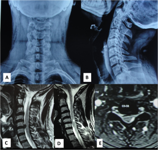
We report a patient with C5-6 cervical disc prolapse that presented with radiculopathy symptoms in the right upper limb, which was refractory to conservative care. He underwent a C5-6 ACDF and reported complete relief from symptoms at 4 weeks. He developed deteriorating symptoms over the next 10 weeks and presented at 14 weeks follow-up with severe myeloradiculopathy symptoms on the left upper limb with upper limb weakness. A fresh MRI identified an intradural extramedullary tumor with cystic changes at the index surgery level. This was treated with tumor excision and histopathology confirmed a diagnosis of schwannoma. Simultaneous presence of cord signal changes with disc herniation obscured the cystic schwannoma which became apparent later on contrast enhanced MRI imaging.
Conclusion
Careful review of preoperative imaging and contrast MRI study may help in diagnosing cystic schwannomas with concomitant cervical disc herniations that have cord signal changes.
中文翻译:
颈椎间盘突出症伪装的颈神经鞘瘤一例报告
介绍
颈椎椎间盘突出症是引起颈椎脊髓神经根病的常见病症之一。颈前路椎间盘切除融合术(ACDF)是其标准治疗方案。硬膜内神经源性肿瘤相对罕见,可表现为脊髓神经根病的特征。放射成像在此类病理的诊断中发挥着重要作用。
案例报告
我们报告了一名 C5-6 颈椎间盘脱垂患者,其右上肢出现神经根病症状,保守治疗难以治愈。他接受了 C5-6 ACDF,并报告在 4 周后症状完全缓解。在接下来的 10 周内,他的症状不断恶化,并在随访 14 周时出现左上肢严重的脊髓神经根病症状,并伴有上肢无力。最新的 MRI 发现硬膜内髓外肿瘤在指数手术水平上出现囊性变化。经过肿瘤切除治疗,组织病理学证实了神经鞘瘤的诊断。脊髓信号变化与椎间盘突出同时存在,掩盖了囊性神经鞘瘤,而囊性神经鞘瘤后来在对比增强 MRI 成像中变得明显。
结论
仔细审查术前影像学和对比 MRI 研究可能有助于诊断囊性神经鞘瘤伴有颈椎间盘突出症(有脊髓信号变化)。


 京公网安备 11010802027423号