Our official English website, www.x-mol.net, welcomes your
feedback! (Note: you will need to create a separate account there.)
Super-resolution microscopy reveals the nanoscale cluster architecture of the DEK protein cancer biomarker
iScience ( IF 4.6 ) Pub Date : 2023-10-19 , DOI: 10.1016/j.isci.2023.108277 Agnieszka Pierzynska-Mach 1 , Alberto Diaspro 1, 2 , Francesca Cella Zanacchi 1, 3, 4
iScience ( IF 4.6 ) Pub Date : 2023-10-19 , DOI: 10.1016/j.isci.2023.108277 Agnieszka Pierzynska-Mach 1 , Alberto Diaspro 1, 2 , Francesca Cella Zanacchi 1, 3, 4
Affiliation

|
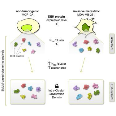
DEK protein, a key chromatin regulator, is strongly overexpressed in various forms of cancer. While conventional microscopy revealed DEK as uniformly distributed within the cell nucleus, advanced super-resolution techniques uncovered cluster-like structures. However, a comprehensive understanding of DEK’s cellular distribution and its implications in cancer and cell growth remained elusive. To bridge this gap, we employed single-molecule localization microscopy (SMLM) to dissect DEK’s nanoscale organization in both normal-like and aggressive breast cancer cell lines. Our investigation included characteristics such as localizations per cluster, cluster areas, and intra-cluster localization densities (ICLDs). We elucidated how cluster features align with different breast cell types and how chromatin decompaction influences DEK clusters in these contexts. Our results indicate that DEK’s intra-cluster localization density and nano-organization remain preserved and not significantly influenced by protein overexpression or chromatin compaction changes. This study advances the understanding of DEK’s role in cancer and underscores its stable nanoscale behavior.
中文翻译:

超分辨率显微镜揭示了 DEK 蛋白质癌症生物标志物的纳米级簇结构
DEK 蛋白是一种关键的染色质调节因子,在各种形式的癌症中强烈过度表达。虽然传统显微镜显示 DEK 均匀分布在细胞核内,但先进的超分辨率技术却发现了簇状结构。然而,对 DEK 的细胞分布及其对癌症和细胞生长的影响的全面了解仍然难以实现。为了弥补这一差距,我们采用单分子定位显微镜 (SMLM) 来剖析 DEK 在正常乳腺癌细胞系和侵袭性乳腺癌细胞系中的纳米级组织。我们的调查包括每个簇的定位、簇区域和簇内定位密度 (ICLD) 等特征。我们阐明了簇特征如何与不同乳腺细胞类型相一致,以及染色质解压缩如何影响这些背景下的 DEK 簇。我们的结果表明,DEK 的簇内定位密度和纳米组织仍然保留,并且不受蛋白质过度表达或染色质压缩变化的显着影响。这项研究增进了对 DEK 在癌症中作用的理解,并强调了其稳定的纳米级行为。
更新日期:2023-10-19
中文翻译:

超分辨率显微镜揭示了 DEK 蛋白质癌症生物标志物的纳米级簇结构
DEK 蛋白是一种关键的染色质调节因子,在各种形式的癌症中强烈过度表达。虽然传统显微镜显示 DEK 均匀分布在细胞核内,但先进的超分辨率技术却发现了簇状结构。然而,对 DEK 的细胞分布及其对癌症和细胞生长的影响的全面了解仍然难以实现。为了弥补这一差距,我们采用单分子定位显微镜 (SMLM) 来剖析 DEK 在正常乳腺癌细胞系和侵袭性乳腺癌细胞系中的纳米级组织。我们的调查包括每个簇的定位、簇区域和簇内定位密度 (ICLD) 等特征。我们阐明了簇特征如何与不同乳腺细胞类型相一致,以及染色质解压缩如何影响这些背景下的 DEK 簇。我们的结果表明,DEK 的簇内定位密度和纳米组织仍然保留,并且不受蛋白质过度表达或染色质压缩变化的显着影响。这项研究增进了对 DEK 在癌症中作用的理解,并强调了其稳定的纳米级行为。


 京公网安备 11010802027423号