European Spine Journal ( IF 2.6 ) Pub Date : 2023-10-07 , DOI: 10.1007/s00586-023-07973-1 Stefan Motov 1, 2 , B Stemmer 2 , P Krauss 2 , C Maurer 3 , E Shiban 2

|
Background
There is only limited data on the management of cerebrospinal fluid (CSF) fistulas after cervical endoscopic spine surgery. We investigated the current literature for treatment options and present a case of a patient who was treated with CT-guided epidural fibrin patch.
Methods
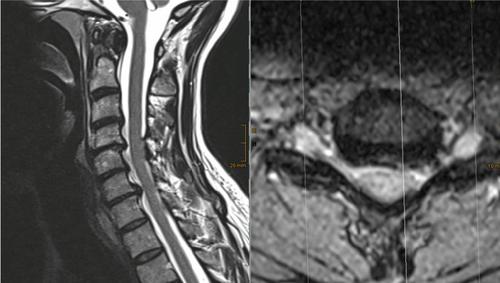
We present the case of a 47-year-old female patient with a suspected CSF fistula after endoscopic decompression for C7 foraminal stenosis. She was readmitted 8 days after surgery with dysesthesia in both upper extremities, orthostatic headache and neck pain, which worsened during mobilization. A CSF leak was suspected on spinal magnetic resonance imaging. A computer tomography (CT)-guided epidural blood patch was performed with short-term relief. A second CT-guided epidural fibrin patch was executed and the patient improved thereafter and was discharged at home without sensorimotor deficits or sequelae. We investigated the current literature for complications after endoscopic spine surgery and for treatment of postoperative CSF fistulas.
Results
Although endoscopic and open revision surgery with dura repair were described in previous studies, dural tears in endoscopic surgery are frequently treated conservatively. In our case, the patient was severely impaired by a persistent CSF fistula. We opted for a less invasive treatment and performed a CT-guided fibrin patch which resulted in a complete resolution of patient’s symptoms.
Discussion and conclusion
CSF fistulas after cervical endoscopic spine procedures are rare complications. Conservative treatment or revision surgery are the standard of care. CT-guided epidural fibrin patch was an efficient and less invasive option in our case.
中文翻译:

使用 CT 引导硬膜外纤维蛋白贴剂进行全内窥镜下颈椎间孔切开术后症状性颈椎脑脊液瘘的治疗
背景
关于颈椎内窥镜脊柱手术后脑脊液 (CSF) 瘘管管理的数据有限。我们调查了目前的治疗方案文献,并介绍了一例接受 CT 引导硬膜外纤维蛋白贴剂治疗的患者。
方法
我们介绍了一名 47 岁女性患者,患者在内窥镜减压后因 C7 椎间孔狭窄而疑似脑脊液瘘。手术后 8 天,她因双上肢感觉迟钝、直立性头痛和颈部疼痛再次入院,活动期间疼痛加重。脊髓磁共振成像怀疑 CSF 泄漏。进行计算机断层扫描 (CT) 引导的硬膜外血贴,短期缓解。执行第二次 CT 引导硬膜外纤维蛋白贴剂,此后患者病情好转,无感觉运动障碍或后遗症在家中出院。我们调查了内窥镜脊柱手术后并发症和术后脑脊液瘘治疗的当前文献。
结果
尽管在以前的研究中描述了内窥镜和开放翻修手术联合硬脑膜修复,但内窥镜手术中的硬脑膜撕裂经常被保守治疗。在我们的案例中,患者因持续性 CSF 瘘管而受到严重损害。我们选择了一种侵入性较小的治疗,并进行了 CT 引导下的纤维蛋白贴剂,导致患者的症状完全消退。
讨论和结论
颈椎内窥镜脊柱手术后的脑脊液瘘管是罕见的并发症。保守治疗或翻修手术是标准护理。在我们的案例中,CT 引导的硬膜外纤维蛋白贴剂是一种有效且侵入性较小的选择。


 京公网安备 11010802027423号