Endocrine ( IF 3.0 ) Pub Date : 2023-09-16 , DOI: 10.1007/s12020-023-03515-3
Jiao Ma 1 , Xiaoyong Wang 2 , Mingsong Tang 2 , Chunyin Zhang 1, 3, 4
|
Objective
To establish a prediction model for preoperatively predicting grade 1 and grade 2/3 tumors in patients with pancreatic neuroendocrine tumors (PNETs) based on 68Ga-DOTATATE PET/CT.
Methods
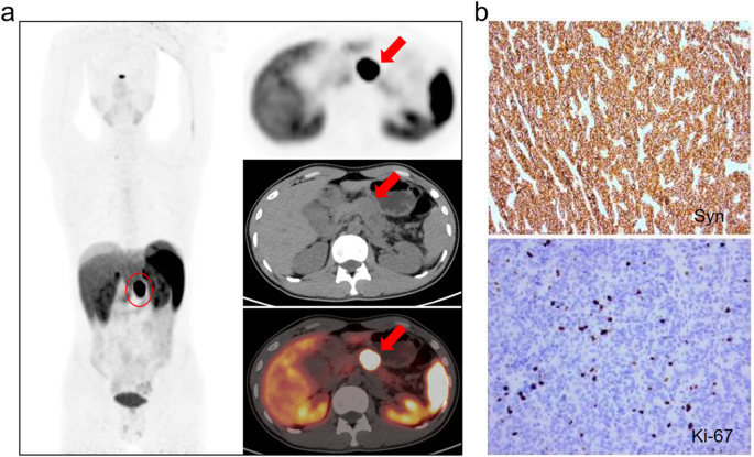
Clinical data of 41 patients with PNETs were included in this study. According to the pathological results, they were divided into grade 1 and grade 2/3. 68Ga-DOTATATE PET/CT images were collected within one month before surgery. The clinical risk factors and significant radiological features were filtered, and a clinical predictive model based on these clinical and radiological features was established. 3D slicer was used to extracted 107 radiomic features from the region of interest (ROI) of 68Ga-dotata PET/CT images. The Pearson correlation coefficient (PCC), recursive feature elimination (REF) based five-fold cross validation were adopted for the radiomic feature selection, and a radiomic score was computed subsequently. The comprehensive model combining the clinical risk factors and the rad-score was established as well as the nomogram. The performance of above clinical model and comprehensive model were evaluated and compared.
Results
Adjacent organ invasion, N staging, and M staging were the risk factors for PNET grading (p < 0.05). 12 optimal radiomic features (3 PET radiomic features, 9 CT radiomic features) were screen out. The clinical predictive model achieved an area under the curve (AUC) of 0.785. The comprehensive model has better predictive performance (AUC = 0.953).
Conclusion
We proposed a comprehensive nomogram model based on 68Ga-DOTATATE PET/CT to predict grade 1 and grade 2/3 of PNETs and assist personalized clinical diagnosis and treatment plans for patients with PNETs.
中文翻译:
基于68Ga-DOTATATE PET/CT的术前胰腺神经内分泌肿瘤分级预测
客观的
建立基于68 Ga-DOTATATE PET/CT 的术前预测胰腺神经内分泌肿瘤(PNET)患者 1 级和 2/3 级肿瘤的预测模型。
方法
本研究纳入了 41 名 PNET 患者的临床数据。根据病理结果分为1级和2/3级。术前1个月内收集68张Ga-DOTATATE PET/CT图像。筛选出临床危险因素和显着的放射学特征,并建立基于这些临床和放射学特征的临床预测模型。 3D 切片器用于从68 个Ga-dotata PET/CT 图像的感兴趣区域 (ROI) 中提取 107 个放射组学特征。采用皮尔逊相关系数(PCC)、基于递归特征消除(REF)的五重交叉验证进行放射组学特征选择,并随后计算放射组学评分。建立了结合临床危险因素和rad评分的综合模型以及列线图。对上述临床模型和综合模型的性能进行了评价和比较。
结果
邻近器官侵犯、N 分期和 M 分期是 PNET 分级的危险因素( p < 0.05)。筛选出 12 个最佳放射组学特征(3 个 PET 放射组学特征,9 个 CT 放射组学特征)。临床预测模型的曲线下面积 (AUC) 为 0.785。综合模型具有更好的预测性能(AUC = 0.953)。
结论
我们提出了基于68 Ga-DOTATATE PET/CT 的综合列线图模型来预测 1 级和 2/3 级 PNET,并协助 PNET 患者制定个性化临床诊断和治疗计划。


 京公网安备 11010802027423号