目的
胞浆内黑色素是透明细胞肉瘤(CCS)的一个特征,它是一种特别致命的软组织肉瘤。 [ 18 F]- N -(2-(二乙氨基)乙基)-5-(2-(2-(2-氟乙氧基)乙氧基)乙氧基)吡啶甲酰胺 ([ 18 F]-PFPN) 是一种正电子发射断层扫描 (PET)探针具有高黑色素亲和力。因此,本研究旨在探讨黑色素靶向[ 18 F]-PFPN PET在CCS患者中的可行性。
方法
这项前瞻性单中心研究招募了经病理证实的 CCS 患者。 [ 18 F]-FDG PET/计算机断层扫描和[ 18 F]-PFPN PET/磁共振成像扫描彼此在1周内进行。病灶数量以及[ 18 F]-FDG和[ 18 F]-PFPN PET参数(最大标准化摄取值[SUVmax]、平均标准化摄取值[SUVmean]、代谢/黑色素肿瘤体积[MTV/MLTV]和总病灶收集糖酵解/黑色素[TLG/TLM])。
结果
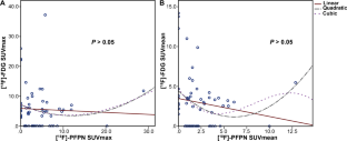
招募了三名 CCS 患者并接受了 PET 成像。 [ 18 F]-PFPN PET共检测到56个病灶,包括原发灶和远处转移灶。在[ 18 F]-PFPN 和[ 18 F]-FDG PET 上未检测到相同的病变。 [ 18 F]-PFPN 上 [ 18 F]-FDG 成像漏检 12 个病灶(12/39,30.77%),[ 18 F]-PFPN 成像漏检 20 个病灶(20/47,42.55%) [ 18 F]-FDG。在定量分析中,所有病灶的[ 18 F]-FDG SUVmean (4.60 ± 3.24) 均高于[ 18 F]-PFPN SUVmean (3.0 ± 2.63) ( P = 0.01)。 [ 18 F]-PFPN 和 [ 18 F]-FDG 的 SUVmax、SUVmean、MLTV/MTV、TLM/TLG 值之间无显着相关性( P > 0.05)。
结论
黑色素靶向[ 18 F]-PFPN PET成像对于CCS的诊断是可行的。 [ 18 F]-PFPN和[ 18 F]-FDG PET成像显示出不同的成像特征,证明了示踪剂的互补作用。 CCS 患者首选联合使用两种成像方式。
临床试验注册
NCT05963035。
 "点击查看英文标题和摘要"
Melanin-targeted [18F]-PFPN PET imaging may shed light for clear cell sarcoma
Purpose
Intracytoplasmic melanin pigment is a characteristic of clear cell sarcoma (CCS), which is a particularly deadly type of soft-tissue sarcoma. [18F]-N-(2-(diethylamino)ethyl)-5-(2-(2-(2-fluoroethoxy)ethoxy)ethoxy)picolinamide ([18F]-PFPN) is a positron emission tomography (PET) probe characterized by high melanin affinity. Therefore, this study aimed to investigate the feasibility of melanin-targeted [18F]-PFPN PET in patients with CCS.
Methods
This prospective single-centre study recruited patients with pathologically confirmed CCS. [18F]-FDG PET/computed tomography and [18F]-PFPN PET/magnetic resonance imaging scans were performed within 1 week of each other. The lesion numbers and [18F]-FDG and [18F]-PFPN PET parameters (maximum standardized uptake value [SUVmax], mean standardized uptake value [SUVmean], metabolic/melanotic tumour volume [MTV/MLTV], and total lesion glycolysis/melanin [TLG/TLM]) were collected.
Results
Three patients with CCS were recruited and received PET imaging. A total of 56 lesions were detected on [18F]-PFPN PET, including primary tumour and distant metastases. Identical lesions were not detected on [18F]-PFPN and [18F]-FDG PET. Twelve lesions (12/39, 30.77%) on [18F]-FDG imaging were missed on [18F]-PFPN, and 20 lesions (20/47, 42.55%) on [18F]-PFPN imaging were missed on [18F]-FDG. In quantitative analysis, the [18F]-FDG SUVmean (4.60 ± 3.24) was higher than the [18F]-PFPN SUVmean (3.0 ± 2.63) in all lesions (P = 0.01). No significant correlations were found between the SUVmax, SUVmean, MLTV/MTV, and TLM/TLG values of [18F]-PFPN and [18F]-FDG (P > 0.05).
Conclusion
Melanin-targeted [18F]-PFPN PET imaging is feasible for the diagnosis of CCS. Different imaging features were displayed on [18F]-PFPN and [18F]-FDG PET imaging, demonstrating the complementary role of the tracers. Combined use of the two imaging modalities would be preferred in patients with CCS.
Clinical Trial Registration
NCT05963035.


 京公网安备 11010802027423号