Scientific Reports ( IF 3.8 ) Pub Date : 2023-09-05 , DOI: 10.1038/s41598-023-41378-w Hongyuan Liu 1 , Zongping Li 1 , Yafei Xue 2 , Tianzhi Zhao 2 , Yingxi Wu 2
|
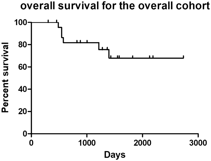
Intracranial chondrosarcoma is a rare tumor with limited reports. We reviewed the clinical outcomes, imaging findings, and pathological characteristics at three centers to improve the diagnosis and treatment of intracranial chondrosarcoma. We retrospectively analyzed 26 patients with intracranial chondrosarcoma who had undergone surgical treatment at Tangdu Hospital of Air Force Military Medical University, Mianyang Central Hospital, and Nanchong Central Hospital from January 2010 to July 2022. Clinical manifestations, imaging features, surgical treatment, prognosis, and overall survival (OS) were analyzed. All 26 chondrosarcomas were located at the skull base. Gross total resection (GTR), subtotal resection (STR), and partial resection (PR) were performed in 14, 10, and 2 cases, respectively. Four cases underwent endoscopic transnasal surgery, while the remaining cases underwent craniotomy. The clinical symptoms were evaluated 1 week after surgery, and 15 cases were relieved to varying degrees. Postoperative complications included pulmonary infection, subcutaneous hydrops, dysphagia and choking, facial numbness, abducens paralysis, and intracranial infection (ICI). Fifteen cases received postoperative adjuvant radiotherapy. Seven cases showed recurrence: two with PR, four with STR, and one with GTR. Six cases received reoperation or radiotherapy after tumor progression, and one untreated patient died 5 months after tumor recurrence. The extent of tumor resection (HR 21.74, 95% CI 1.25–376.6, P = 0.03) and pathological grading (HR 131.99, 95% CI 4.05–4300.5, P = 0.006) were associated with improved OS. We presented our experience in the treatment of intracranial chondrosarcoma at three centers in the past 12 years. Intracranial chondrosarcoma lacked typical imaging features and are difficult to differentiate from other skull base lesions. Maximum extent of tumor resection with minimal injury to neurological function remains the most important treatment strategy. The extent of surgical resection and pathological grading were found to be predictors for OS.
中文翻译:
26例颅内软骨肉瘤临床结局的多中心回顾性分析
颅内软骨肉瘤是一种罕见的肿瘤,报道有限。我们回顾了三个中心的临床结果、影像学表现和病理特征,以改进颅内软骨肉瘤的诊断和治疗。我们回顾性分析2010年1月至2022年7月在空军军医大学唐都医院、绵阳市中心医院、南充市中心医院接受手术治疗的26例颅内软骨肉瘤患者的临床表现、影像学特征、手术治疗、预后及治疗情况。分析总生存期(OS)。所有 26 个软骨肉瘤均位于颅底。共行大体全切除(GTR)、次全切除(STR)、部分切除(PR),分别为14例、10例、2例。4例接受内窥镜经鼻手术,其余病例接受开颅手术。术后1周评估临床症状,15例均有不同程度缓解。术后并发症包括肺部感染、皮下积水、吞咽困难和窒息、面部麻木、外展肌麻痹和颅内感染(ICI)。15例接受术后辅助放疗。7 例复发:2 例 PR,4 例 STR,1 例 GTR。6例患者在肿瘤进展后接受了再次手术或放疗,1例未经治疗的患者在肿瘤复发后5个月死亡。肿瘤切除范围(HR 21.74,95% CI 1.25-376.6,P = 0.03)和病理分级(HR 131.99,95% CI 4.05-4300.5,P = 0.006)与 OS 改善相关。我们介绍了过去12年来我们在三个中心治疗颅内软骨肉瘤的经验。颅内软骨肉瘤缺乏典型的影像学特征,难以与其他颅底病变鉴别。最大限度地切除肿瘤并尽量减少对神经功能的损伤仍然是最重要的治疗策略。研究发现手术切除范围和病理分级是 OS 的预测因素。


 京公网安备 11010802027423号