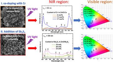
我们报告了两种增强 ZnO:Ca 系统中近红外发射的新策略。在第一种策略中,我们采用湿化学法合成了 ZnO:Ca 纳米粒子,并掺杂了不同浓度的 Cr。SEM 图像显示,ZnO:Ca,Cr 纳米颗粒具有颗粒状形貌,当 Cr 浓度从 4 mol% 增加到 17 mol% 后,其尺寸从 33 nm 增加到 110 nm。X射线衍射(XRD)分析表明,Cr含量为4 mol%的ZnO:Ca,Cr样品具有纯六方相,而Cr浓度高于11 mol%合成的其他样品则呈现六方相和少量的六方相。 Cr 2 O 3含量(<3%)。此外,这些样品的光致发光(PL)光谱显示出蓝色和近红外区域的两个主要发射峰(λexc = 265 和 365 nm)。特别是,由 17 mol% Cr 制成的 ZnO:Ca,Cr 样品在 700-850 nm 区域具有最强的 NIR 发射,并且只有该样品在 805 nm 处显示出显着的 NIR 发射峰。此外,CIE 图显示 ZnO:Ca,Cr 样品的发射在 265 nm 激发下位于蓝色区域,但在 365 nm 激发下被调整到橙色区域。在第二种策略中,在ZnO:Ca的合成过程中添加不同量的Sb 2 S 3以形成ZnO:Ca/Sb 2 S 3复合材料。在这种情况下,我们获得了不同形态的Sb 2 S 3晶粒,棒状和片状含量分别为10、28和34wt%。此外,复合材料呈现出六方相和斜方相的混合物,分别对应于ZnO和Sb 2 S 3。ZnO:Ca/Sb 2 S 3复合材料在650-800 nm区域具有近红外发射峰,并且其强度随着Sb 2 S 3含量的增加而增加。此外,根据 CIE 地图,它们的可见光发射从蓝色调整为橙红色或冷白光。总体而言,最佳 ZnO:Ca,Cr 样品的排放量比 ZnO:Ca 参考样品和最佳 ZnO:Ca/Sb 2 S 3 高 312%和37 %复合材料(在 265 nm 激发下),分别。总的来说,XPS 研究表明存在氧空位缺陷,从而在所有样品中产生可见光-近红外发射。这里提出的 NIR 发射增强策略在低成本下是可行的,并且这里产生的 NIR 发射发生在第一个生物窗口(700-1000 nm)内,这对于生物医学应用很有意义。
 "点击查看英文标题和摘要"
Novel strategies for the enhancement of the NIR emission in the Ca doped ZnO system: Doping with Cr or adding Sb2S3
We report two novel strategies to enhance the NIR emission in the ZnO:Ca system. In the first strategy, we synthesized ZnO:Ca nanoparticles using a wet chemical method and doped them with various concentration of Cr. SEM images showed that the ZnO:Ca,Cr nanoparticles had a grain-like morphology and their sizes increased from 33 to 110 nm after incrementing the Cr concentration from 4 to 17 mol%. The analysis by X-ray diffraction (XRD) revealed that he ZnO:Ca,Cr sample made with 4 mol% of Cr had a pure hexagonal phase but the other samples synthesized with Cr concentration above 11 mol% presented the hexagonal phase and a small content of Cr2O3 (<3%). Moreover, the photoluminescence (PL) spectra of these samples showed two main emission peaks in the blue and NIR regions (λexc = 265 and 365 nm). In particular, the ZnO:Ca,Cr sample made with 17 mol% of Cr had the strongest NIR emission in the 700–850 nm region and only this sample showed a prominent NIR emission peak at 805 nm. Moreover, the CIE maps showed that the emissions of the ZnO:Ca,Cr samples are located in the blue region under excitation at 265 nm, but it was tuned to the orange region under excitation at 365 nm. In the second strategy, different amounts of Sb2S3 was added during the synthesis of ZnO:Ca to form ZnO:Ca/Sb2S3 composites. In this case, we obtained different morphologies of grains, rod-like and sheet-like for Sb2S3 contents of 10, 28 and 34 wt%, respectively. In addition, the composites presented a mixture of hexagonal and orthorhombic phases, which corresponded to the ZnO and Sb2S3, respectively. The ZnO:Ca/Sb2S3 composites had NIR emissions peaks in the 650–800 nm region and their intensity increased with the content of Sb2S3. Furthermore, their visible emission was tuned from blue to orange-red or to cold white light according to the CIE maps. Overall, the best ZnO:Ca,Cr sample had 312% and 37% higher emission than the ZnO:Ca reference sample and the best ZnO:Ca/Sb2S3 composite (under 265 nm excitation), respectively. In general, XPS studies showed the existence of oxygen vacancy defects, which produced the visible-NIR emissions in all the samples. The strategies presented here for NIR emission enhancement are feasible at low cost and the NIR emissions produced here occur within the first biological window (700–1000 nm), which is of interest for biomedical applications.


 京公网安备 11010802027423号