Neurosurgical Review ( IF 2.5 ) Pub Date : 2023-07-29 , DOI: 10.1007/s10143-023-02097-y
Hu Sun 1 , Hui Zhang 2 , Lijie Jing 1 , Hao Zhao 1 , Bing Chen 3 , Wei Song 4
|
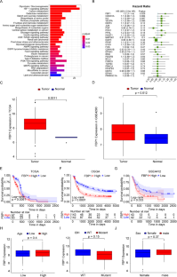
Hypoxia has been shown to contribute to tumor immunosuppressive microenvironment and is an effective prognostic indicator. This study aimed to screen prognostic hypoxia-related genes (HRGs) in glioblastoma and investigate the association between HRGs and tumor immunosuppressive microenvironment. The glioblastoma-related mRNA data were collected from TCGA, GEO, and CGGA databases. Totally 200 HRGs were obtained from the GSEA website. The prognostic HRGs were screened by univariate Cox regression analysis. Somatic mutation data of glioblastoma from TCGA was visualized using the “maftools” of R package. Immune cell infiltration proportions were calculated by CIBERSORT. The TISIDB online tool was applied to analyze the relationship between HRGs and immunoinhibitors as well as the HRG expression in different glioblastoma immune and molecular subtypes. Hub gene’s mRNA and protein levels in cell lines were determined by qRT-PCR and western blot, respectively. The effects of hub gene knockdown on cell viability and migration ability were evaluated employing CCK8 and wound healing assays. The univariate Cox regression showed that high level of FBP1 (fructose-1,6-bisphosphatase 1) was a poor prognostic biomarker, and FBP1 was mainly expressed in lymphocyte depleted immune subtype of glioblastoma. High FBP1 mRNA and protein levels have been successfully validated in vitro. The somatic mutation analysis suggested that TP53 mutation rate was the highest in the high FBP1 glioblastoma group, while EGFR mutation rate was the highest in the low FBP1 glioblastoma group. In the high FBP1 group, the infiltration proportions and types of immune cells were less, dominated by macrophages M2, and the expression of CTLA4, LAG3, TIGIT, PDL1, and PDL2 was significantly upregulated. The expression of FBP1 was positively correlated with several immunoinhibitors, such as IL-10 and TGFβ-1. In conclusion, we demonstrated that FBP1 could serve as a prognostic biomarker for glioblastoma. The immune microenvironment in the high FBP1 group might be suppressed by up-regulating immune checkpoints and immunoinhibitors.
中文翻译:
FBP1 是一种潜在的预后生物标志物,与胶质母细胞瘤的肿瘤免疫抑制微环境相关
缺氧已被证明有助于肿瘤免疫抑制微环境,并且是有效的预后指标。本研究旨在筛选胶质母细胞瘤中的预后缺氧相关基因(HRG)并研究HRG与肿瘤免疫抑制微环境之间的关联。胶质母细胞瘤相关 mRNA 数据收集自 TCGA、GEO 和 CGGA 数据库。从 GSEA 网站总共获得了 200 个 HRG。通过单变量 Cox 回归分析筛选预后 HRG。使用 R 包的“maftools”可视化 TCGA 中胶质母细胞瘤的体细胞突变数据。通过CIBERSORT计算免疫细胞浸润比例。应用TISIDB在线工具分析HRG与免疫抑制剂的关系以及HRG在不同胶质母细胞瘤免疫和分子亚型中的表达。分别通过qRT-PCR和蛋白质印迹测定细胞系中Hub基因的mRNA和蛋白水平。采用 CCK8 和伤口愈合测定评估 hub 基因敲低对细胞活力和迁移能力的影响。单变量 Cox 回归显示,高水平的 FBP1(果糖-1,6-双磷酸酶 1)是预后不良的生物标志物,FBP1 主要表达于淋巴细胞耗竭的免疫亚型胶质母细胞瘤中。高 FBP1 mRNA 和蛋白质水平已在体外得到成功验证。体细胞突变分析提示,高FBP1胶质母细胞瘤组TP53突变率最高,而低FBP1胶质母细胞瘤组EGFR突变率最高。 高FBP1组免疫细胞浸润比例和类型较少,以巨噬细胞M2为主,CTLA4、LAG3、TIGIT、PDL1、PDL2表达显着上调。 FBP1的表达与IL-10、TGFβ-1等多种免疫抑制剂呈正相关。总之,我们证明 FBP1 可以作为胶质母细胞瘤的预后生物标志物。高FBP1组的免疫微环境可能通过上调免疫检查点和免疫抑制剂而受到抑制。


 京公网安备 11010802027423号