Our official English website, www.x-mol.net, welcomes your
feedback! (Note: you will need to create a separate account there.)
Cover Image, Volume 71, Issue 9
Glia ( IF 5.4 ) Pub Date : 2023-07-04 , DOI: 10.1002/glia.24210
Linjuan Feng , Hsuan Lo , Zhaoxiang Hong , Jiahao Zheng , Yuhong Yan , Zucheng Ye , Xiaochun Chen , Xiaodong Pan
Glia ( IF 5.4 ) Pub Date : 2023-07-04 , DOI: 10.1002/glia.24210
Linjuan Feng , Hsuan Lo , Zhaoxiang Hong , Jiahao Zheng , Yuhong Yan , Zucheng Ye , Xiaochun Chen , Xiaodong Pan
|
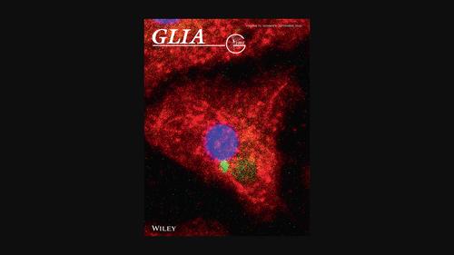
Cover Illustration: Confocal laser scanning microscopy image of triple staining with Iba1 (red), CX3CR1 (green) and DAPI (blue) in the primary microglia of Nfatc1fl/fl; Lyz2Cre/+ mice at 4 h after α-Synuclein inducement. (See Feng, L, et al. https://doi.org/10.1002/glia.24422)
中文翻译:

封面图片,第 71 卷,第 9 期
封面插图:Nfatc1 fl/fl 原代小胶质细胞中 Iba1(红色)、CX3CR1(绿色)和 DAPI(蓝色)三重染色的共焦激光扫描显微镜图像;α-突触核蛋白诱导后 4 小时的Lyz2 Cre/+小鼠。(参见 Feng, L 等人。https://doi.org/10.1002/glia.24422)
更新日期:2023-07-08
中文翻译:
封面图片,第 71 卷,第 9 期
封面插图:Nfatc1 fl/fl 原代小胶质细胞中 Iba1(红色)、CX3CR1(绿色)和 DAPI(蓝色)三重染色的共焦激光扫描显微镜图像;α-突触核蛋白诱导后 4 小时的Lyz2 Cre/+小鼠。(参见 Feng, L 等人。https://doi.org/10.1002/glia.24422)


 京公网安备 11010802027423号