Our official English website, www.x-mol.net, welcomes your
feedback! (Note: you will need to create a separate account there.)
High-precision, low-complexity, high-resolution microscopy-based cell sorting
Lab on a Chip ( IF 6.1 ) Pub Date : 2023-06-07 , DOI: 10.1039/d3lc00242j Tobias Gerling 1, 2 , Neus Godino 1 , Felix Pfisterer 1 , Nina Hupf 1 , Michael Kirschbaum 1
Lab on a Chip ( IF 6.1 ) Pub Date : 2023-06-07 , DOI: 10.1039/d3lc00242j Tobias Gerling 1, 2 , Neus Godino 1 , Felix Pfisterer 1 , Nina Hupf 1 , Michael Kirschbaum 1
Affiliation

|
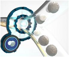
Continuous flow cell sorting based on image analysis is a powerful concept that exploits spatially-resolved features in cells, such as subcellular protein localisation or cell and organelle morphology, to isolate highly specialised cell types that were previously inaccessible to biomedical research, biotechnology, and medicine. Recently, sorting protocols have been proposed that achieve impressive throughput by combining ultra-high flow rates with sophisticated imaging and data processing protocols. However, moderate image quality and high complex experimental setups still prevent the full potential of image-activated cell sorting from being a general-purpose tool. Here, we present a new low-complexity microfluidic approach based on high numerical aperture wide-field microscopy and precise dielectrophoretic cell handling. It provides high-quality images with unprecedented resolution in image-activated cell sorting (i.e., 216 nm). In addition, it also allows long image processing times of several hundred milliseconds for thorough image analysis, while ensuring reliable and low-loss cell processing. Using our approach, we sorted live T cells based on subcellular localisation of fluorescence signals and demonstrated that purities above 80% are possible while targeting maximum yields and sample volume throughputs in the range of μl min−1. We were able to recover 85% of the target cells analysed. Finally, we ensure and quantify the full vitality of the sorted cells cultivating the cells for a period of time and through colorimetric viability tests.
中文翻译:

高精度、低复杂性、高分辨率的基于显微镜的细胞分选
基于图像分析的连续流式细胞分选是一个强大的概念,它利用细胞中的空间分辨特征,例如亚细胞蛋白质定位或细胞和细胞器形态,来分离以前生物医学研究、生物技术和医学无法获得的高度专业化的细胞类型。最近,人们提出了通过将超高流速与复杂的成像和数据处理协议相结合来实现令人印象深刻的吞吐量的分选协议。然而,中等的图像质量和高度复杂的实验设置仍然阻碍了图像激活细胞分选作为通用工具的全部潜力。在这里,我们提出了一种基于高数值孔径宽视场显微镜和精确介电泳细胞处理的新的低复杂性微流体方法。即,216 nm)。此外,它还允许数百毫秒的长图像处理时间,以进行彻底的图像分析,同时确保可靠和低损耗的细胞处理。使用我们的方法,我们根据荧光信号的亚细胞定位对活 T 细胞进行分类,并证明纯度可以高于 80%,同时目标是最大产量和 μl min -1范围内的样品体积吞吐量。我们能够回收 85% 的分析靶细胞。最后,我们通过培养细胞一段时间并通过比色活力测试来确保和量化分选细胞的全部活力。
更新日期:2023-06-07
中文翻译:

高精度、低复杂性、高分辨率的基于显微镜的细胞分选
基于图像分析的连续流式细胞分选是一个强大的概念,它利用细胞中的空间分辨特征,例如亚细胞蛋白质定位或细胞和细胞器形态,来分离以前生物医学研究、生物技术和医学无法获得的高度专业化的细胞类型。最近,人们提出了通过将超高流速与复杂的成像和数据处理协议相结合来实现令人印象深刻的吞吐量的分选协议。然而,中等的图像质量和高度复杂的实验设置仍然阻碍了图像激活细胞分选作为通用工具的全部潜力。在这里,我们提出了一种基于高数值孔径宽视场显微镜和精确介电泳细胞处理的新的低复杂性微流体方法。即,216 nm)。此外,它还允许数百毫秒的长图像处理时间,以进行彻底的图像分析,同时确保可靠和低损耗的细胞处理。使用我们的方法,我们根据荧光信号的亚细胞定位对活 T 细胞进行分类,并证明纯度可以高于 80%,同时目标是最大产量和 μl min -1范围内的样品体积吞吐量。我们能够回收 85% 的分析靶细胞。最后,我们通过培养细胞一段时间并通过比色活力测试来确保和量化分选细胞的全部活力。


 京公网安备 11010802027423号