当前位置:
X-MOL 学术
›
ACS Chem. Neurosci.
›
论文详情
Our official English website, www.x-mol.net, welcomes your
feedback! (Note: you will need to create a separate account there.)
Identifying Neural Signatures of Dopamine Signaling with Machine Learning
ACS Chemical Neuroscience ( IF 4.1 ) Pub Date : 2023-06-02 , DOI: 10.1021/acschemneuro.3c00001 Siamak K Sorooshyari 1 , Nicholas Ouassil 2 , Sarah J Yang 2 , Markita P Landry 2, 3, 4, 5
ACS Chemical Neuroscience ( IF 4.1 ) Pub Date : 2023-06-02 , DOI: 10.1021/acschemneuro.3c00001 Siamak K Sorooshyari 1 , Nicholas Ouassil 2 , Sarah J Yang 2 , Markita P Landry 2, 3, 4, 5
Affiliation

|
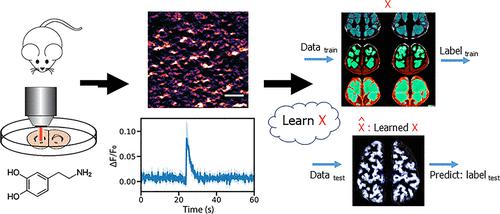
The emergence of new tools to image neurotransmitters, neuromodulators, and neuropeptides has transformed our understanding of the role of neurochemistry in brain development and cognition, yet analysis of this new dimension of neurobiological information remains challenging. Here, we image dopamine modulation in striatal brain tissue slices with near-infrared catecholamine nanosensors (nIRCat) and implement machine learning to determine which features of dopamine modulation are unique to changes in stimulation strength, and to different neuroanatomical regions. We trained a support vector machine and a random forest classifier to decide whether the recordings were made from the dorsolateral striatum (DLS) versus the dorsomedial striatum (DMS) and find that machine learning is able to accurately distinguish dopamine release that occurs in DLS from that occurring in DMS in a manner unachievable with canonical statistical analysis. Furthermore, our analysis determines that dopamine modulatory signals including the number of unique dopamine release sites and peak dopamine released per stimulation event are most predictive of neuroanatomy. This is in light of integrated neuromodulator amount being the conventional metric used to monitor neuromodulation in animal studies. Lastly, our study finds that machine learning discrimination of different stimulation strengths or neuroanatomical regions is only possible in adult animals, suggesting a high degree of variability in dopamine modulatory kinetics during animal development. Our study highlights that machine learning could become a broadly utilized tool to differentiate between neuroanatomical regions or between neurotypical and disease states, with features not detectable by conventional statistical analysis.
中文翻译:

通过机器学习识别多巴胺信号的神经特征
对神经递质、神经调节剂和神经肽进行成像的新工具的出现改变了我们对神经化学在大脑发育和认知中的作用的理解,但对神经生物学信息这一新维度的分析仍然具有挑战性。在这里,我们使用近红外儿茶酚胺纳米传感器(nIRCat)对纹状体脑组织切片中的多巴胺调节进行成像,并实施机器学习来确定多巴胺调节的哪些特征对于刺激强度的变化以及不同的神经解剖区域是独特的。我们训练了支持向量机和随机森林分类器来确定记录是从背外侧纹状体(DLS)还是背内侧纹状体(DMS)进行的,并发现机器学习能够准确地区分 DLS 中发生的多巴胺释放在 DMS 中以典型统计分析无法实现的方式发生。此外,我们的分析确定多巴胺调节信号(包括独特的多巴胺释放位点的数量和每次刺激事件释放的多巴胺峰值)最能预测神经解剖学。这是因为综合神经调节剂的量是动物研究中用于监测神经调节的传统指标。最后,我们的研究发现,机器学习对不同刺激强度或神经解剖区域的区分只有在成年动物中才有可能,这表明动物发育过程中多巴胺调节动力学存在高度可变性。我们的研究强调,机器学习可能成为一种广泛使用的工具,用于区分神经解剖区域或神经典型状态和疾病状态,其特征无法通过传统统计分析检测到。
更新日期:2023-06-02
中文翻译:

通过机器学习识别多巴胺信号的神经特征
对神经递质、神经调节剂和神经肽进行成像的新工具的出现改变了我们对神经化学在大脑发育和认知中的作用的理解,但对神经生物学信息这一新维度的分析仍然具有挑战性。在这里,我们使用近红外儿茶酚胺纳米传感器(nIRCat)对纹状体脑组织切片中的多巴胺调节进行成像,并实施机器学习来确定多巴胺调节的哪些特征对于刺激强度的变化以及不同的神经解剖区域是独特的。我们训练了支持向量机和随机森林分类器来确定记录是从背外侧纹状体(DLS)还是背内侧纹状体(DMS)进行的,并发现机器学习能够准确地区分 DLS 中发生的多巴胺释放在 DMS 中以典型统计分析无法实现的方式发生。此外,我们的分析确定多巴胺调节信号(包括独特的多巴胺释放位点的数量和每次刺激事件释放的多巴胺峰值)最能预测神经解剖学。这是因为综合神经调节剂的量是动物研究中用于监测神经调节的传统指标。最后,我们的研究发现,机器学习对不同刺激强度或神经解剖区域的区分只有在成年动物中才有可能,这表明动物发育过程中多巴胺调节动力学存在高度可变性。我们的研究强调,机器学习可能成为一种广泛使用的工具,用于区分神经解剖区域或神经典型状态和疾病状态,其特征无法通过传统统计分析检测到。


 京公网安备 11010802027423号