Micron ( IF 2.5 ) Pub Date : 2023-03-22 , DOI: 10.1016/j.micron.2023.103448 Arash Rabbani 1 , Masoud Babaei 2 , Masoumeh Gharib 3

|
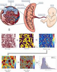
In this study, a novel method of data augmentation has been presented for the segmentation of placental histological images when the labeled data are scarce. This method generates new realizations of the placenta intervillous morphology while maintaining the general textures and orientations. As a result, a diversified artificial dataset of images is generated that can be used for training deep learning segmentation models. We have observed that on average the presented method of data augmentation led to a 42% decrease in the binary cross-entropy loss of the validation dataset compared to the common approach in the literature. Additionally, the morphology of the intervillous space is studied under the effect of the proposed image reconstruction technique, and the diversity of the artificially generated population is quantified. We have demonstrated that the proposed method results in a more accurate morphological characterization of the placental intervillous space with an average relative error of 6.5%, which is significantly lower than the 11.5% error observed with conventional augmentation techniques. Due to the high resemblance of the generated images to the real ones, applications of the proposed method may not be limited to placental histological images, and it is recommended that other types of tissue be investigated in future studies.
中文翻译:

基于单个标记图像的胎盘绒毛间隙的自动分割和形态学表征
在这项研究中,提出了一种新的数据增强方法,用于在标记数据稀缺时分割胎盘组织学图像。这种方法产生了胎盘绒毛间形态的新认识,同时保持了一般的纹理和方向。结果,生成了多样化的人工图像数据集,可用于训练深度学习分割模型。我们观察到,与文献中的常用方法相比,所提出的数据增强方法平均导致验证数据集的二元交叉熵损失减少 42%。此外,在所提出的图像重建技术的作用下研究了绒毛间隙的形态,并对人工生成的种群的多样性进行了量化。我们已经证明,所提出的方法可以更准确地对胎盘绒毛间隙进行形态学表征,平均相对误差为 6.5%,显着低于传统增强技术观察到的 11.5% 误差。由于生成的图像与真实图像高度相似,所提出方法的应用可能不仅限于胎盘组织学图像,建议在未来的研究中研究其他类型的组织。


 京公网安备 11010802027423号