Nature ( IF 50.5 ) Pub Date : 2023-03-08 , DOI: 10.1038/s41586-023-05788-0 Xiaoying Chen 1 , Maria Firulyova 2 , Melissa Manis 1 , Jasmin Herz 3, 4 , Igor Smirnov 3, 4 , Ekaterina Aladyeva 3 , Chanung Wang 1 , Xin Bao 1 , Mary Beth Finn 1 , Hao Hu 1 , Irina Shchukina 3 , Min Woo Kim 3, 4 , Carla M Yuede 1 , Jonathan Kipnis 1, 3, 4 , Maxim N Artyomov 3 , Jason D Ulrich 1 , David M Holtzman 1, 4
|
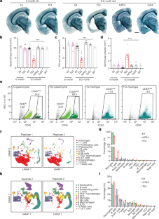
Extracellular deposition of amyloid-β as neuritic plaques and intracellular accumulation of hyperphosphorylated, aggregated tau as neurofibrillary tangles are two of the characteristic hallmarks of Alzheimer’s disease1,2. The regional progression of brain atrophy in Alzheimer’s disease highly correlates with tau accumulation but not amyloid deposition3,4,5, and the mechanisms of tau-mediated neurodegeneration remain elusive. Innate immune responses represent a common pathway for the initiation and progression of some neurodegenerative diseases. So far, little is known about the extent or role of the adaptive immune response and its interaction with the innate immune response in the presence of amyloid-β or tau pathology6. Here we systematically compared the immunological milieux in the brain of mice with amyloid deposition or tau aggregation and neurodegeneration. We found that mice with tauopathy but not those with amyloid deposition developed a unique innate and adaptive immune response and that depletion of microglia or T cells blocked tau-mediated neurodegeneration. Numbers of T cells, especially those of cytotoxic T cells, were markedly increased in areas with tau pathology in mice with tauopathy and in the Alzheimer’s disease brain. T cell numbers correlated with the extent of neuronal loss, and the cells dynamically transformed their cellular characteristics from activated to exhausted states along with unique TCR clonal expansion. Inhibition of interferon-γ and PDCD1 signalling both significantly ameliorated brain atrophy. Our results thus reveal a tauopathy- and neurodegeneration-related immune hub involving activated microglia and T cell responses, which could serve as therapeutic targets for preventing neurodegeneration in Alzheimer’s disease and primary tauopathies.
中文翻译:
小胶质细胞介导的 T 细胞浸润驱动 tau 蛋白病的神经退行性变
β 淀粉样蛋白在细胞外沉积为神经斑,细胞内过度磷酸化、聚集的 tau 蛋白在细胞内堆积为神经原纤维缠结,这是阿尔茨海默病的两个特征1,2 。阿尔茨海默病脑萎缩的区域进展与 tau 积累高度相关,但与淀粉样蛋白沉积无关3,4,5 ,并且 tau 介导的神经退行性变的机制仍然难以捉摸。先天免疫反应是一些神经退行性疾病发生和进展的常见途径。到目前为止,人们对适应性免疫反应的程度或作用及其在β淀粉样蛋白或tau蛋白病理存在下与先天免疫反应的相互作用知之甚少6 。在这里,我们系统地比较了小鼠大脑中淀粉样蛋白沉积或 tau 蛋白聚集和神经变性的免疫环境。我们发现,患有 tau 蛋白病的小鼠(而非患有淀粉样蛋白沉积的小鼠)产生了独特的先天性和适应性免疫反应,并且小胶质细胞或 T 细胞的消耗阻止了 tau 蛋白介导的神经变性。在患有 tau 病的小鼠和患有阿尔茨海默病的大脑中,tau 病理区域的 T 细胞数量,尤其是细胞毒性 T 细胞的数量显着增加。 T 细胞数量与神经元损失程度相关,并且细胞动态地将其细胞特征从激活状态转变为耗尽状态,并伴随着独特的 TCR 克隆扩张。抑制干扰素-γ 和 PDCD1 信号传导均能显着改善脑萎缩。 因此,我们的结果揭示了一个与tau蛋白病和神经变性相关的免疫中枢,涉及激活的小胶质细胞和T细胞反应,这可以作为预防阿尔茨海默病和原发性tau蛋白病中神经变性的治疗靶点。


 京公网安备 11010802027423号