当前位置:
X-MOL 学术
›
Adv. Funct. Mater.
›
论文详情
Our official English website, www.x-mol.net, welcomes your
feedback! (Note: you will need to create a separate account there.)
Integrated and Bifunctional Bilayer 3D Printing Scaffold for Osteochondral Defect Repair
Advanced Functional Materials ( IF 18.5 ) Pub Date : 2023-02-28 , DOI: 10.1002/adfm.202214158 Cairong Li 1, 2 , Wei Zhang 1, 2 , Yangyi Nie 1, 2 , Dongchun Jiang 1, 2 , Jingyi Jia 1, 2 , Wenjing Zhang 1, 2 , Long Li 1, 2 , Zhenyu Yao 1, 2 , Ling Qin 3, 4 , Yuxiao Lai 1, 2, 3
Advanced Functional Materials ( IF 18.5 ) Pub Date : 2023-02-28 , DOI: 10.1002/adfm.202214158 Cairong Li 1, 2 , Wei Zhang 1, 2 , Yangyi Nie 1, 2 , Dongchun Jiang 1, 2 , Jingyi Jia 1, 2 , Wenjing Zhang 1, 2 , Long Li 1, 2 , Zhenyu Yao 1, 2 , Ling Qin 3, 4 , Yuxiao Lai 1, 2, 3
Affiliation

|
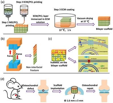
Bioinspired scaffolds with two distinct regions resembling stratified anatomical architecture provide potential strategies for osteochondral defect repair and are studied in preclinical animals. However, delamination of the two layers often causes tissue disjunction between the regenerated cartilage and subchondral bone, leading to few commercially available clinical applications. This study develops an integrated poly(ε-caprolactone) (PCL)-based scaffold for repairing osteochondral defects. An extracellular matrix (ECM)-incorporated 3D printing composite scaffold (ECM/PCL) coated with ECM hydrogel (E-co-E/PCL) is fabricated as the upper layer, and magnesium oxide nanoparticles coated with polydopamine (MgO@PDA)-incorporated composite scaffold (MD/PCL) is fabricated using 3D printing as the bottom layer. The physicochemical and mechanical properties of the bilayer scaffold meet the requirements in designing and fabricating the osteochondral scaffold, especially a strong interface possessed between the two layers. By in vitro study, the integrated scaffold stimulates proliferation, chondrogenic differentiation, and osteogenic differentiation of human bone mesenchymal stem cells. Moreover, the integrated bilayer scaffold exhibits well repair ability to facilitate simultaneous regeneration of cartilage and subchondral bone after implanting into the osteochondral defect in rats. In addition, cartilage “tidemarks” completely regenerated after 12 weeks of implantation of the bilayer scaffold, which indicates no tissue disjunctions formed between the regenerated cartilage and subchondral bone.
中文翻译:

用于骨软骨缺损修复的集成双功能双层 3D 打印支架
具有两个类似于分层解剖结构的不同区域的仿生支架为骨软骨缺损修复提供了潜在的策略,并在临床前动物中进行了研究。然而,两层的分层经常导致再生软骨和软骨下骨之间的组织分离,导致很少有商业上可用的临床应用。本研究开发了一种基于聚(ε-己内酯)(PCL)的集成支架,用于修复骨软骨缺损。将细胞外基质 (ECM) 结合的 3D 打印复合支架 (ECM/PCL) 涂有 ECM 水凝胶 (E-co-E/PCL) 作为上层,并用氧化镁纳米粒子涂有聚多巴胺 (MgO@PDA)-复合支架 (MD/PCL) 是使用 3D 打印作为底层制造的。双层支架的物理化学和力学性能满足骨软骨支架设计和制造的要求,特别是两层之间具有强界面。通过体外研究,集成支架可刺激人骨髓间充质干细胞的增殖、软骨形成分化和成骨分化。此外,集成双层支架具有良好的修复能力,可在植入大鼠骨软骨缺损后促进软骨和软骨下骨的同时再生。此外,在植入双层支架 12 周后,软骨“潮痕”完全再生,这表明再生软骨和软骨下骨之间没有形成组织分离。
更新日期:2023-02-28
中文翻译:

用于骨软骨缺损修复的集成双功能双层 3D 打印支架
具有两个类似于分层解剖结构的不同区域的仿生支架为骨软骨缺损修复提供了潜在的策略,并在临床前动物中进行了研究。然而,两层的分层经常导致再生软骨和软骨下骨之间的组织分离,导致很少有商业上可用的临床应用。本研究开发了一种基于聚(ε-己内酯)(PCL)的集成支架,用于修复骨软骨缺损。将细胞外基质 (ECM) 结合的 3D 打印复合支架 (ECM/PCL) 涂有 ECM 水凝胶 (E-co-E/PCL) 作为上层,并用氧化镁纳米粒子涂有聚多巴胺 (MgO@PDA)-复合支架 (MD/PCL) 是使用 3D 打印作为底层制造的。双层支架的物理化学和力学性能满足骨软骨支架设计和制造的要求,特别是两层之间具有强界面。通过体外研究,集成支架可刺激人骨髓间充质干细胞的增殖、软骨形成分化和成骨分化。此外,集成双层支架具有良好的修复能力,可在植入大鼠骨软骨缺损后促进软骨和软骨下骨的同时再生。此外,在植入双层支架 12 周后,软骨“潮痕”完全再生,这表明再生软骨和软骨下骨之间没有形成组织分离。


 京公网安备 11010802027423号