Our official English website, www.x-mol.net, welcomes your
feedback! (Note: you will need to create a separate account there.)
Ultrasensitive Multiplex Imaging of Cell Surface Proteins via Core-Shell Surface-Enhanced Raman Scattering Nanoprobes
ACS Sensors ( IF 8.2 ) Pub Date : 2023-02-27 , DOI: 10.1021/acssensors.3c00100 Jin Wang 1, 2 , Zheng Tan 3 , Chengcheng Zhu 1 , Li Xu 1 , Xing-Hua Xia 2 , Chen Wang 1, 2
ACS Sensors ( IF 8.2 ) Pub Date : 2023-02-27 , DOI: 10.1021/acssensors.3c00100 Jin Wang 1, 2 , Zheng Tan 3 , Chengcheng Zhu 1 , Li Xu 1 , Xing-Hua Xia 2 , Chen Wang 1, 2
Affiliation

|
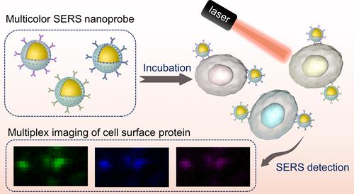
Cell surface proteins, as important components of biological membranes, cover a wide range of important markers of diseases and even cancers. In this regard, precise detection of their expression levels is of crucial importance for both cancer diagnosis and the development of responsive therapeutic strategies. Herein, a size-controlled core-shell Au@ Copper(II) benzene-1,3,5-tricarboxylate (Au@Cu-BTC) nanomaterial was synthesized for specific and simultaneous imaging of multiple protein expression levels on cell membranes. The porous shell of Cu-BTC constructed on Au nanoparticles enabled effective loading of Raman reporter molecules, followed by further modification of the targeting moieties, which equipped the nanoprobe with good specificity and stability. Additionally, given the flexibility of the types of Raman reporter molecules available for loading, the nanoprobes were also demonstrated with good multichannel imaging capabilities. Ultimately, the present strategy of electromagnetic and chemical dual Raman scattering enhancement was successfully applied for the simultaneous detection of varied proteins on cell surfaces with high sensitivity and accuracy. The proposed nanomaterial holds promising applications in biosensing and therapeutic fields, which could not only provide a general strategy for the synthesis of metal–organic framework-based core-shell surface-enhanced Raman scattering nanoprobes but also enable further utilization in multitarget and multichannel cell imaging.
中文翻译:

通过核壳表面增强拉曼散射纳米探针对细胞表面蛋白进行超灵敏多重成像
细胞表面蛋白作为生物膜的重要组成部分,涵盖了广泛的疾病甚至癌症的重要标志物。在这方面,精确检测它们的表达水平对于癌症诊断和响应性治疗策略的发展都至关重要。在此,合成了一种尺寸可控的核-壳 Au@铜 (II) 苯-1,3,5-三羧酸盐 (Au@Cu-BTC) 纳米材料,用于对细胞膜上的多种蛋白质表达水平进行特异性和同步成像。在Au纳米粒子上构建的Cu-BTC多孔壳能够有效负载拉曼报告分子,随后进一步修饰靶向部分,使纳米探针具有良好的特异性和稳定性。此外,鉴于可用于加载的拉曼报告分子类型的灵活性,纳米探针也被证明具有良好的多通道成像能力。最终,本发明的电磁和化学双重拉曼散射增强策略成功应用于以高灵敏度和准确性同时检测细胞表面的各种蛋白质。拟议的纳米材料在生物传感和治疗领域具有广阔的应用前景,不仅可以为基于金属有机骨架的核壳表面增强拉曼散射纳米探针的合成提供通用策略,还可以进一步用于多靶点和多通道细胞成像. 目前的电磁和化学双拉曼散射增强策略已成功应用于以高灵敏度和准确性同时检测细胞表面的各种蛋白质。拟议的纳米材料在生物传感和治疗领域具有广阔的应用前景,不仅可以为基于金属有机骨架的核壳表面增强拉曼散射纳米探针的合成提供通用策略,还可以进一步用于多靶点和多通道细胞成像. 目前的电磁和化学双拉曼散射增强策略已成功应用于以高灵敏度和准确性同时检测细胞表面的各种蛋白质。拟议的纳米材料在生物传感和治疗领域具有广阔的应用前景,不仅可以为基于金属有机骨架的核壳表面增强拉曼散射纳米探针的合成提供通用策略,还可以进一步用于多靶点和多通道细胞成像.
更新日期:2023-02-27
中文翻译:

通过核壳表面增强拉曼散射纳米探针对细胞表面蛋白进行超灵敏多重成像
细胞表面蛋白作为生物膜的重要组成部分,涵盖了广泛的疾病甚至癌症的重要标志物。在这方面,精确检测它们的表达水平对于癌症诊断和响应性治疗策略的发展都至关重要。在此,合成了一种尺寸可控的核-壳 Au@铜 (II) 苯-1,3,5-三羧酸盐 (Au@Cu-BTC) 纳米材料,用于对细胞膜上的多种蛋白质表达水平进行特异性和同步成像。在Au纳米粒子上构建的Cu-BTC多孔壳能够有效负载拉曼报告分子,随后进一步修饰靶向部分,使纳米探针具有良好的特异性和稳定性。此外,鉴于可用于加载的拉曼报告分子类型的灵活性,纳米探针也被证明具有良好的多通道成像能力。最终,本发明的电磁和化学双重拉曼散射增强策略成功应用于以高灵敏度和准确性同时检测细胞表面的各种蛋白质。拟议的纳米材料在生物传感和治疗领域具有广阔的应用前景,不仅可以为基于金属有机骨架的核壳表面增强拉曼散射纳米探针的合成提供通用策略,还可以进一步用于多靶点和多通道细胞成像. 目前的电磁和化学双拉曼散射增强策略已成功应用于以高灵敏度和准确性同时检测细胞表面的各种蛋白质。拟议的纳米材料在生物传感和治疗领域具有广阔的应用前景,不仅可以为基于金属有机骨架的核壳表面增强拉曼散射纳米探针的合成提供通用策略,还可以进一步用于多靶点和多通道细胞成像. 目前的电磁和化学双拉曼散射增强策略已成功应用于以高灵敏度和准确性同时检测细胞表面的各种蛋白质。拟议的纳米材料在生物传感和治疗领域具有广阔的应用前景,不仅可以为基于金属有机骨架的核壳表面增强拉曼散射纳米探针的合成提供通用策略,还可以进一步用于多靶点和多通道细胞成像.


 京公网安备 11010802027423号