当前位置:
X-MOL 学术
›
ACS Chem. Neurosci.
›
论文详情
Our official English website, www.x-mol.net, welcomes your
feedback! (Note: you will need to create a separate account there.)
In Vivo and 3D Imaging Technique(s) for Spatiotemporal Mapping of Pathological Events in Experimental Model(s) of Spinal Cord Injury
ACS Chemical Neuroscience ( IF 4.1 ) Pub Date : 2023-02-14 , DOI: 10.1021/acschemneuro.2c00643 Divya Goyal 1 , Hemant Kumar 1
ACS Chemical Neuroscience ( IF 4.1 ) Pub Date : 2023-02-14 , DOI: 10.1021/acschemneuro.2c00643 Divya Goyal 1 , Hemant Kumar 1
Affiliation

|
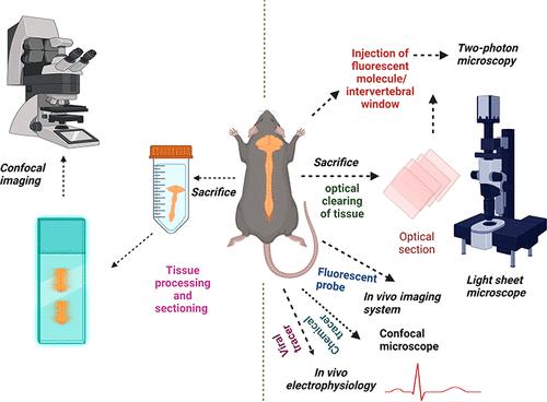
Endothelial damage, astrogliosis, microgliosis, and neuronal degeneration are the most common events after spinal cord injury (SCI). Studies highlighted that studying the spatiotemporal profile of these events might provide a deeper understanding of the pathophysiology of SCI. For imaging of these events, available conventional techniques such as 2-dimensional histology and immunohistochemistry (IHC) are well established and frequently used to visualize and detect the altered expression of the protein of interest involved in these events. However, the technique requires the physical sectioning of the tissue, and results are also open to misinterpretation. Currently, researchers are focusing more attention toward the advanced tools for imaging the spinal cord’s various physiological and pathological parameters. The tools include two-photon imaging, light sheet fluorescence microscopy, in vivo imaging system with fluorescent probes, and in vivo chemical and fluorescent protein-expressing viral-tracers. These techniques outperform the limitations associated with conventional techniques in various aspects, such as optical sectioning of tissue, 3D reconstructed imaging, and imaging of particular planes of interest. In addition to this, these techniques are minimally invasive and less time-consuming. In this review, we will discuss the various advanced imaging methodologies that will evolve in the future to explore the fundamental mechanisms after SCI.
中文翻译:

脊髓损伤实验模型中病理事件时空映射的体内和 3D 成像技术
内皮损伤、星形胶质细胞增生、小胶质细胞增生和神经元变性是脊髓损伤 (SCI) 后最常见的事件。研究强调,研究这些事件的时空特征可能会加深对 SCI 病理生理学的理解。对于这些事件的成像,可用的常规技术,如二维组织学和免疫组织化学 (IHC) 已经很好地建立起来,并经常用于可视化和检测这些事件中所涉及的感兴趣蛋白质的表达改变。然而,该技术需要对组织进行物理切片,而且结果也容易被误解。目前,研究人员更加关注用于对脊髓各种生理和病理参数进行成像的先进工具。这些工具包括双光子成像,带有荧光探针的体内成像系统,以及体内化学和荧光蛋白表达病毒示踪剂。这些技术在各个方面都优于传统技术的局限性,例如组织的光学切片、3D 重建成像和特定感兴趣平面的成像。除此之外,这些技术是微创且耗时较少的。在这篇综述中,我们将讨论未来将发展的各种先进成像方法,以探索 SCI 后的基本机制。
更新日期:2023-02-14
中文翻译:

脊髓损伤实验模型中病理事件时空映射的体内和 3D 成像技术
内皮损伤、星形胶质细胞增生、小胶质细胞增生和神经元变性是脊髓损伤 (SCI) 后最常见的事件。研究强调,研究这些事件的时空特征可能会加深对 SCI 病理生理学的理解。对于这些事件的成像,可用的常规技术,如二维组织学和免疫组织化学 (IHC) 已经很好地建立起来,并经常用于可视化和检测这些事件中所涉及的感兴趣蛋白质的表达改变。然而,该技术需要对组织进行物理切片,而且结果也容易被误解。目前,研究人员更加关注用于对脊髓各种生理和病理参数进行成像的先进工具。这些工具包括双光子成像,带有荧光探针的体内成像系统,以及体内化学和荧光蛋白表达病毒示踪剂。这些技术在各个方面都优于传统技术的局限性,例如组织的光学切片、3D 重建成像和特定感兴趣平面的成像。除此之外,这些技术是微创且耗时较少的。在这篇综述中,我们将讨论未来将发展的各种先进成像方法,以探索 SCI 后的基本机制。


 京公网安备 11010802027423号