Progress in Nuclear Magnetic Resonance Spectroscopy ( IF 7.3 ) Pub Date : 2023-02-10 , DOI: 10.1016/j.pnmrs.2023.02.002 Jacob Chen Ming Low 1 , Alan J Wright 1 , Friederike Hesse 1 , Jianbo Cao 1 , Kevin M Brindle 1

|
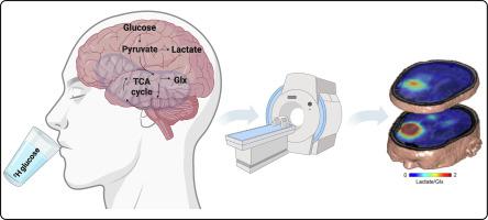
Deuterium metabolic imaging (DMI) is an emerging clinically-applicable technique for the non-invasive investigation of tissue metabolism. The generally short T1 values of 2H-labeled metabolites in vivo can compensate for the relatively low sensitivity of detection by allowing rapid signal acquisition in the absence of significant signal saturation. Studies with deuterated substrates, including [6,6'-2H2]glucose, [2H3]acetate, [2H9]choline and [2,3-2H2]fumarate have demonstrated the considerable potential of DMI for imaging tissue metabolism and cell death in vivo. The technique is evaluated here in comparison with established metabolic imaging techniques, including PET measurements of 2-deoxy-2-[18F]fluoro-D-glucose (FDG) uptake and 13C MR imaging of the metabolism of hyperpolarized 13C-labeled substrates.
中文翻译:

使用氘标记底物的代谢成像
氘代谢成像(DMI)是一种新兴的临床适用技术,用于组织代谢的非侵入性研究。体内2 H标记代谢物的通常较短的T 1值可以通过允许在不存在显着信号饱和的情况下快速采集信号来补偿相对较低的检测灵敏度。对氘化底物(包括[6,6'- 2 H 2 ]葡萄糖、[ 2 H 3 ]乙酸盐、[ 2 H 9 ]胆碱和[2,3- 2 H 2 ]富马酸盐)的研究表明,DMI 在以下方面具有巨大的潜力:体内组织代谢和细胞死亡成像。此处将该技术与已建立的代谢成像技术进行比较进行评估,包括 2-脱氧-2-[ 18 F]氟-D-葡萄糖 (FDG) 摄取的 PET 测量和超极化13 C 标记代谢的13 C MR 成像基材。


 京公网安备 11010802027423号