Analytica Chimica Acta ( IF 5.7 ) Pub Date : 2023-02-06 , DOI: 10.1016/j.aca.2023.340949 David Vicente-Zurdo 1 , Beatriz Gómez-Gómez 1 , Iván Romero-Sánchez 1 , Noelia Rosales-Conrado 1 , María Eugenia León-González 1 , Yolanda Madrid 1

|
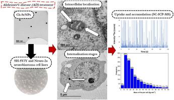
Alzheimer's disease (AD) is the most prevalent neurodegenerative disease, representing 80% of the total dementia cases. The “amyloid cascade hypothesis” stablishes that the aggregation of the beta-amyloid protein (Aβ42) is the first event that subsequently triggers AD development. Selenium nanoparticles stabilized with chitosan (Ch-SeNPs) have demonstrated excellent anti-amyloidogenic properties in previous works, leading to an improvement of AD aetiology. Here, the in vitro effect of selenium species in AD model cell line has been study to obtain a better assessment of their effects in AD treatment. For this purpose, mouse neuroblastoma (Neuro-2a) and human neuroblastoma (SH-SY5Y) cell lines were used. Cytotoxicity of selenium species, such as selenomethionine (SeMet), Se-methylselenocysteine (MeSeCys) and Ch-SeNPs, has been determined by 3-(4,5-Dimethylthiazol-2-yl)-2,5-diphenyltetrazolium bromide (MTT) and flow cytometry methods. Intracellular localisation of Ch-SeNPs, and their pathway through SH-SY5Y cell line, have been evaluated by transmission electron microscopy (TEM). The uptake and accumulation of selenium species by both neuroblastoma cell lines have been quantified at single cell level by single cell- Inductively Coupled Plasma with Mass Spectrometry detection (SC-ICP-MS), with a previous optimisation of transport efficiency using gold nanoparticles (AuNPs) ((69 ± 3) %) and 2.5 mm calibration beads ((92 ± 8) %). Results showed that Ch-SeNPs would be more readily accumulated by both cell lines than organic species being accumulation ranges between 1.2 and 89.5 fg Se cell−1 for Neuro-2a and 3.1–129.8 fg Se cell−1 for SH-SY5Y exposed to 250 μM Ch-SeNPs. Data obtained were statistically treated using chemometric tools. These results provide an important insight into the interaction of Ch-SeNPs with neuronal cells, which could support their potential use in AD treatment.
中文翻译:

通过使用细胞毒性测定、TEM 和单细胞-ICP-MS 对与阿尔茨海默氏病相关的神经母细胞瘤细胞系中硒纳米粒子和其他硒物种的细胞毒性、摄取和积累
阿尔茨海默病 (AD) 是最普遍的神经退行性疾病,占痴呆病例总数的 80%。“淀粉样蛋白级联假说”证实,β-淀粉样蛋白 (Aβ 42 ) 的聚集是随后触发 AD 发展的第一个事件。用壳聚糖 (Ch-SeNPs) 稳定的硒纳米粒子在以前的工作中表现出优异的抗淀粉样蛋白生成特性,从而改善了 AD 病因学。在这里,体外已经研究了硒种类在 AD 模型细胞系中的作用,以便更好地评估它们在 AD 治疗中的作用。为此,使用了小鼠神经母细胞瘤 (Neuro-2a) 和人神经母细胞瘤 (SH-SY5Y) 细胞系。已通过 3-(4,5-二甲基噻唑-2-基)-2,5-二苯基四唑溴化物 (MTT) 测定了硒物种(例如硒代甲硫氨酸 (SeMet)、硒-甲基硒代半胱氨酸 (MeSeCys) 和 Ch-SeNPs)的细胞毒性和流式细胞术方法。Ch-SeNP 的细胞内定位及其通过 SH-SY5Y 细胞系的途径已通过透射电子显微镜 (TEM) 进行了评估。两种神经母细胞瘤细胞系对硒物质的摄取和积累已通过单细胞电感耦合等离子体质谱检测 (SC-ICP-MS) 在单细胞水平上进行了量化,先前使用金纳米粒子 (AuNP) ((69 ± 3) %) 和 2.5 mm 校准珠 ((92 ± 8) %) 优化了传输效率。结果表明,Ch-SeNPs 比有机物种更容易被两种细胞系积累,积累范围在 1.2 和 89.5 fg Se 细胞之间-1对于 Neuro-2a 和 3.1–129.8 fg Se 细胞-1对于暴露于 250 μM Ch-SeNPs 的 SH-SY5Y。使用化学计量学工具对获得的数据进行统计处理。这些结果为 Ch-SeNPs 与神经元细胞的相互作用提供了重要的见解,这可能支持它们在 AD 治疗中的潜在用途。


 京公网安备 11010802027423号