当前位置:
X-MOL 学术
›
Environ. Sci.: Nano
›
论文详情
Our official English website, www.x-mol.net, welcomes your
feedback! (Note: you will need to create a separate account there.)
Fate, uptake and gut toxicity of two colloidal silver products in mice: how micro X-ray fluorescence, micro X-ray absorption spectroscopy and near-infrared spectroscopy provide new insights in food nanotoxicology
Environmental Science: Nano ( IF 5.8 ) Pub Date : 2023-01-24 , DOI: 10.1039/d2en01135b Kevin Gillois 1 , Camille Rivard 2, 3 , Cecile Levasseur-Garcia 4 , Valerie Bezirard 1 , Helene Terrisse 5 , Renaud Leonard 6 , Catherine Robbe-Masselot 6 , Emmanuelle Maguin 7 , Mathias L. Richard 7, 8 , Vassilia Theodorou 1 , Marie-Helene Ropers 9 , Muriel Mercier-Bonin 1, 10 , Herve Robert 1
Environmental Science: Nano ( IF 5.8 ) Pub Date : 2023-01-24 , DOI: 10.1039/d2en01135b Kevin Gillois 1 , Camille Rivard 2, 3 , Cecile Levasseur-Garcia 4 , Valerie Bezirard 1 , Helene Terrisse 5 , Renaud Leonard 6 , Catherine Robbe-Masselot 6 , Emmanuelle Maguin 7 , Mathias L. Richard 7, 8 , Vassilia Theodorou 1 , Marie-Helene Ropers 9 , Muriel Mercier-Bonin 1, 10 , Herve Robert 1
Affiliation
|
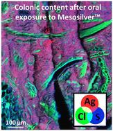
Silver biodistribution and gut toxicity of two commercially available colloidal silver products, Mesosilver™ and AgC, were evaluated in male mice. AgC is composed solely of ionic silver (Ag+) while Mesosilver™ contains a mix of silver nanoparticles (AgNPs) and Ag+ ions. After high-dose (approximately 3 mg per kg body weight (bw) per day) sub-chronic exposure, silver accumulation was close for Mesosilver™ and AgC. The combination of micro X-ray fluorescence and micro X-ray absorption spectroscopy showed that metallic AgNPs and Ag+ ions initially contained in Mesosilver™ were subjected to physicochemical modifications during their fate in the gut. In the colon, most Ag atoms were oxidized and dissolved to form Ag complexes with thiol groups (–SH) of proteins and/or peptides. Sub-chronic exposure at lower dose (150 μg per kg bw per day) led to a moderate impact on the gut barrier for both colloidal silver products. An increase in colonic LCN-2 was observed only after AgC exposure. For gut microbiota at the genus level, exposure to Mesosilver™ led to a decrease in Ruminococcus and Anaerosporobacter, while Intestinimonas increased. Exposure to AgC resulted in an increase in Clostridium sp. ASF356 and Tyzzerella, while the relative abundance of Anaerosporobacter decreased. In addition, the Saccharomycetes fungal population increased. Near-infrared spectroscopy was able to satisfactorily discriminate the Mesosilver™- vs. AgC-exposed mice for both exposure doses. This study highlights the applicability of biophysics-based methodologies for providing novel insights into colloidal silver uptake, fate and toxicological effects after oral exposure.
中文翻译:

两种胶体银产品在小鼠体内的命运、摄取和肠道毒性:微 X 射线荧光、微 X 射线吸收光谱和近红外光谱如何为食品纳米毒理学提供新见解
在雄性小鼠中评估了两种市售胶体银产品 Mesosilver™ 和 AgC 的银生物分布和肠道毒性。AgC 仅由离子银 (Ag + ) 组成,而 Mesosilver™ 包含银纳米粒子 (AgNP) 和 Ag +离子的混合物。在高剂量(每天每公斤体重 (bw) 大约 3 毫克)亚慢性暴露后,Mesosilver™ 和 AgC 的银积累接近。结合显微 X 射线荧光和显微 X 射线吸收光谱表明,金属 AgNPs 和 Ag +最初包含在 Mesosilver™ 中的离子在它们在肠道中的命运过程中受到了物理化学修饰。在结肠中,大多数 Ag 原子被氧化并溶解,与蛋白质和/或肽的硫醇基 (–SH) 形成 Ag 络合物。较低剂量(每天每千克体重 150 微克)的亚慢性接触对两种胶体银产品的肠道屏障产生中度影响。仅在接触 AgC 后才观察到结肠 LCN-2 的增加。对于属水平的肠道微生物群,暴露于 Mesosilver™ 导致瘤胃球菌和厌氧孢子杆菌减少,而肠单胞菌增加。暴露于 AgC 导致梭状芽孢杆菌的增加。ASF356 和Tyzzerella,而厌氧孢子杆菌的相对丰度下降。此外,酵母菌种群数量增加。近红外光谱能够令人满意地区分两种暴露剂量的 Mesosilver™和AgC 暴露小鼠。本研究强调了基于生物物理学的方法论的适用性,为口服暴露后胶体银的摄取、命运和毒理学效应提供新的见解。
更新日期:2023-01-24
中文翻译:
两种胶体银产品在小鼠体内的命运、摄取和肠道毒性:微 X 射线荧光、微 X 射线吸收光谱和近红外光谱如何为食品纳米毒理学提供新见解
在雄性小鼠中评估了两种市售胶体银产品 Mesosilver™ 和 AgC 的银生物分布和肠道毒性。AgC 仅由离子银 (Ag + ) 组成,而 Mesosilver™ 包含银纳米粒子 (AgNP) 和 Ag +离子的混合物。在高剂量(每天每公斤体重 (bw) 大约 3 毫克)亚慢性暴露后,Mesosilver™ 和 AgC 的银积累接近。结合显微 X 射线荧光和显微 X 射线吸收光谱表明,金属 AgNPs 和 Ag +最初包含在 Mesosilver™ 中的离子在它们在肠道中的命运过程中受到了物理化学修饰。在结肠中,大多数 Ag 原子被氧化并溶解,与蛋白质和/或肽的硫醇基 (–SH) 形成 Ag 络合物。较低剂量(每天每千克体重 150 微克)的亚慢性接触对两种胶体银产品的肠道屏障产生中度影响。仅在接触 AgC 后才观察到结肠 LCN-2 的增加。对于属水平的肠道微生物群,暴露于 Mesosilver™ 导致瘤胃球菌和厌氧孢子杆菌减少,而肠单胞菌增加。暴露于 AgC 导致梭状芽孢杆菌的增加。ASF356 和Tyzzerella,而厌氧孢子杆菌的相对丰度下降。此外,酵母菌种群数量增加。近红外光谱能够令人满意地区分两种暴露剂量的 Mesosilver™和AgC 暴露小鼠。本研究强调了基于生物物理学的方法论的适用性,为口服暴露后胶体银的摄取、命运和毒理学效应提供新的见解。


 京公网安备 11010802027423号