CardioVascular and Interventional Radiology ( IF 2.8 ) Pub Date : 2022-12-07 , DOI: 10.1007/s00270-022-03325-6
Daniel H Kwak 1 , Qian Yu 1 , Mira Malavia 2 , Emily Sellers 1 , Adam Said 3 , Mikin Patel 1 , Divya Kumari 1 , Osman Ahmed 1
|
Purpose
To investigate risk factors associated with post-microwave ablation (MWA) abscess development.
Materials and Methods
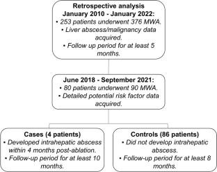
A retrospective case–control analysis was conducted to identify hepatic MWA performed at a single tertiary medical center between January 2010 and January 2022. Case and control patients were defined as those who did or did not develop intrahepatic abscess within 3 months following MWA, respectively. Correlations between risk factors and post-MWA abscess development were assessed by Fisher’s exact test.
Results
Between 2010 and 2022, 253 patients underwent 376 MWA sessions with post-ablation abscess complication rate of 1.1% (4/376). Complications associated with intrahepatic abscess included bacteremia, empyema, pleural abscess, subcutaneous abscess, cholangitis, bile leak, biliocutaneous and arterio-biliary fistulae, and pseudoaneurysm. One patient expired from septic shock 5 days post-ablation. All abscesses were treated by percutaneous drainage and antibiotics. One patient required concomitant placement of a biliary stent and embolization of a biliocutaneous tract. History of Sphincter of Oddi manipulation (p < 0.01), cholangiocarcinoma (p < 0.05), transarterial radioembolization (TARE) to the index lesion (p < 0.05), and abnormal serum alkaline phosphatase levels (p < 0.05) were significantly correlated with post-MWA abscess. The risk of developing post-MWA abscesses for patients with a history of cholangiocarcinoma or a history of Sphincter of Oddi manipulation were 20.0% and 27.2%, respectively.
Conclusion
Patients with prior Sphincter of Oddi manipulation, cholangiocarcinoma, or TARE are at greater risk of developing post-MWA abscess.
中文翻译:
肝肿瘤经皮微波消融治疗后脓肿发生的危险因素
目的
调查与微波消融后 (MWA) 脓肿发展相关的风险因素。
材料和方法
进行了一项回顾性病例对照分析,以确定 2010 年 1 月至 2022 年 1 月期间在一家三级医疗中心进行的肝脏 MWA。病例和对照患者分别定义为在 MWA 后 3 个月内发生或未发生肝内脓肿的患者。通过 Fisher 精确检验评估风险因素与 MWA 后脓肿发展之间的相关性。
结果
2010 年至 2022 年间,253 名患者接受了 376 次 MWA 治疗,消融后脓肿并发症发生率为 1.1% (4/376)。与肝内脓肿相关的并发症包括菌血症、脓胸、胸膜脓肿、皮下脓肿、胆管炎、胆漏、胆皮瘘和动胆瘘以及假性动脉瘤。一名患者在消融后 5 天死于感染性休克。所有脓肿均通过经皮引流和抗生素治疗。一名患者需要同时放置胆道支架和栓塞胆道皮道。Oddi 括约肌操作史 ( p < 0.01)、胆管癌 ( p < 0.05)、指标病变经动脉放射栓塞术 (TARE) ( p < 0.05) 和异常血清碱性磷酸酶水平 ( p < 0.05) 与 MWA 后脓肿显着相关。有胆管癌病史或 Oddi 括约肌操作病史的患者发生 MWA 后脓肿的风险分别为 20.0% 和 27.2%。
结论
既往有 Oddi 括约肌操作、胆管癌或 TARE 病史的患者发生 MWA 后脓肿的风险更大。


 京公网安备 11010802027423号