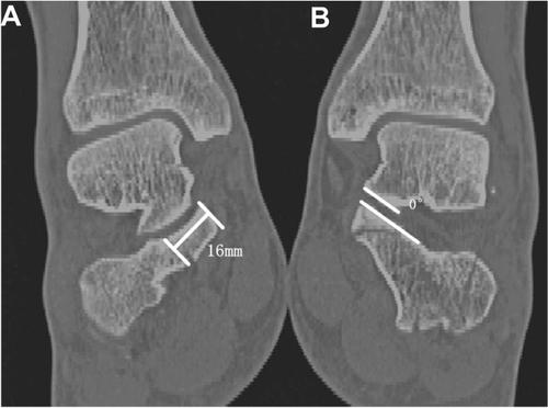
距骨托由骨间韧带和三角韧带复合体紧紧束缚在距骨上,被认为是“恒定的碎片”。然而,缺乏对支持部分的解剖模式的研究。因此,本研究旨在通过在二维 (2D) 和三维 (3D) 条件下应用计算机断层扫描 (CT) 映射来确定跟骨关节内骨折 (ICF) 中支持性骨折的发生率和移位情况. 从 2019 年 1 月到 2020 年 12 月,回顾性评估了 67 名患者的 81 个 ICF 的 CT 图像,此外,还记录了患者的基本人口统计学和损伤机制。支持性骨折的患病率在矢状或冠状 CT 平面上具有特征。半脱位,成角,并且注意到支撑骨部分的平移。通过减少重建的骨折碎片以适应 sustentaculum tali 的模型,生成了 3D 地图。总体而言,81 例 ICF 中有 21 例 (25.9%) 发生支持性骨折,15 例 (71.4%) 无移位,6 例 (28.6%) 移位,平均冠状角为 21.9°,无粉碎性骨折。71 例 (87.7%) 的 sustentaculum tali 和距骨之间的关系在解剖学上对齐,10 例 (12.3%) 的半脱位。根据研究,3D映射显示,大多数骨折线从大腿后突前部开始,斜向延伸至拇长屈肌腱沟。此外,本研究详细描述了 ICF 中支持性碎片的频率(移位或关节脱位)。这一发现反驳了支撑片的“恒定”理论,因为支撑片的粉碎性骨折很少见。这些骨折模式的专业知识可能会影响固定概念和手术方法的进展。此外,我们进一步推测,支撑碎片的位移更有可能出现在高阶桑德斯分类中。然而,需要更大的样本量来进一步验证这一立场。我们进一步推测,在高阶 Sanders 分类中出现支撑碎片的位移的可能性要大得多。然而,需要更大的样本量来进一步验证这一立场。我们进一步推测,在高阶 Sanders 分类中出现支撑碎片的位移的可能性要大得多。然而,需要更大的样本量来进一步验证这一立场。
 "点击查看英文标题和摘要"
Two and three-dimensional CT mapping of the sustentacular fragment in intra-articular calcaneal fractures
The sustentaculum tali are tightly bound to the talus by the interosseous and deltoid ligament complex and have been considered a ‘‘constant fragment”. Yet there is a dearth of study on the anatomical patterns of the sustentacular segment. Consequently, this study is designated with the purpose of defining the prevalence and displacement of sustentacular fractures in intra-articular calcaneal fractures (ICFs) applying computed tomography (CT) mapping in both two-dimensional (2D) and three-dimensional (3D) conditions. From January 2019 to December 2020, the CT images of sixty-seven patients with eighty-one ICFs were retrospectively evaluated, besides, basic patient demographics and mechanisms of injury were documented. And the prevalence of sustentacular fractures was characterized in the sagittal or coronal CT planes. The subluxation, angulation, and translation of the portion of the sustentacular bone were noted. By decreasing rebuilt fracture fragments to suit a model of the sustentaculum tali, a 3D map was generated. Overall, the sustentacular fracture in 21 (25.9%) of the 81 ICFs, 15 (71.4%) were nondisplaced, 6 (28.6%) were displaced, and mean coronal angulation was 21.9°, and no comminuted. The relationship between sustentaculum tali and the talus was anatomically aligned in 71 (87.7%), and subluxation in 10 (12.3%). According to the research, 3D mapping demonstrated that most fracture lines start from the anterior of the sustentaculum tali, extending obliquely to the sulcus of the flexor halluces longus tendon. Moreover, this study provides a detailed description (displacement or articular dislocation) of the frequency of sustentacular fragments in ICFs. The finding disproves the ‘‘constant’’ theory of the sustentacular fragments, due to the fact that comminuted fracture of sustentaculum tali was rare. And the expertise of these fracture patterns may affect the progress of fixation concepts and surgical approaches. Moreover, we further speculated that the displacement of the sustentacular fragment was considerably more probable to emerge in the higher-order Sanders classification. Nevertheless, bigger sample size is required to further validate this position.


 京公网安备 11010802027423号