Brain Imaging and Behavior ( IF 2.4 ) Pub Date : 2022-11-21 , DOI: 10.1007/s11682-022-00742-6
Gita Thapaliya 1 , Sally Eldeghaidy 2, 3, 4 , Michael Asghar 3 , Jordan McGing 2, 3 , Shellie Radford 2 , Susan Francis 2, 3 , Gordon William Moran 2, 3, 5
|
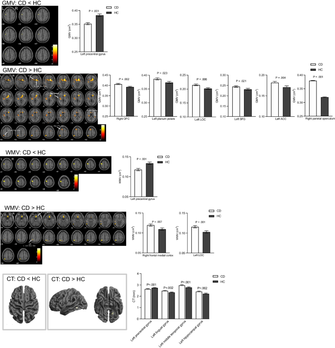
Alterations in grey matter volume (GMV) and cortical thickness (CT) in Crohn’s disease (CD) patients has been previously documented. However, the findings are inconsistent, and not a true representation of CD burden, as only CD patients in remission have been studied thus far. We investigate alterations in brain morphometry in patients with active CD and those in remission, and study relationships between brain structure and key symptoms of fatigue, abdominal pain, and extraintestinal manifestations (EIM). Magnetic Resonance Imaging brain scans were collected in 89 participants; 34 CD participants with active disease, 13 CD participants in remission and 42 healthy controls (HCs); Voxel based morphometry (VBM) assessed GMV and white matter volume (WMV), and surface-based analysis assessed cortical thickness (CT). We show a significant reduction in global cerebrospinal fluid (CSF) volume in CD participants compared with HCs, as well as, a reduction in regional GMV, WMV and CT in the left precentral gyrus (motor cortex), and an increase in GMV in the frontal brain regions in CD compared with HCs. Atrophy of the supplementary motor area (SMA) was associated with greater fatigue in CD. We also show alterations in brain structure in multiple regions in CD associated with abdominal pain and extraintestinal inflammations (EIMs). These brain structural alterations likely reflect neuroplasticity to a chronic systemic inflammatory response, abdominal pain, EIMs and fatigue. These findings will aid our understanding of the cross-linking between chronic inflammation, brain structural changes and key unexplained CD symptomatology like fatigue.
中文翻译:
克罗恩病中枢神经系统形态变化与主要症状的关系
克罗恩病 (CD) 患者的灰质体积 (GMV) 和皮质厚度 (CT) 的变化先前已有记载。然而,这些发现是不一致的,并且不是 CD 负担的真实代表,因为迄今为止只研究了处于缓解状态的 CD 患者。我们调查活动性 CD 患者和缓解期患者脑形态计量学的改变,并研究脑结构与疲劳、腹痛和肠外表现 (EIM) 等主要症状之间的关系。收集了 89 名参与者的磁共振成像脑部扫描;34 名患有活动性疾病的 CD 参与者、13 名处于缓解期的 CD 参与者和 42 名健康对照者 (HC);基于体素的形态测量学 (VBM) 评估 GMV 和白质体积 (WMV),基于表面的分析评估皮质厚度 (CT)。我们显示,与 HC 相比,CD 参与者的整体脑脊液 (CSF) 体积显着减少,左侧中央前回(运动皮层)的区域 GMV、WMV 和 CT 减少,并且 GMV 增加与 HC 相比,CD 中的额叶脑区。辅助运动区 (SMA) 的萎缩与 CD 患者的疲劳程度有关。我们还展示了与腹痛和肠外炎症 (EIM) 相关的 CD 多个区域的大脑结构改变。这些脑结构改变可能反映了对慢性全身炎症反应、腹痛、EIM 和疲劳的神经可塑性。这些发现将有助于我们理解慢性炎症、大脑结构变化和关键的无法解释的 CD 症状(如疲劳)之间的交叉联系。


 京公网安备 11010802027423号