当前位置:
X-MOL 学术
›
ACS Appl. Mater. Interfaces
›
论文详情
Our official English website, www.x-mol.net, welcomes your
feedback! (Note: you will need to create a separate account there.)
Anthracene Single-Crystal Scintillators for Computer Tomography Scanning
ACS Applied Materials & Interfaces ( IF 8.3 ) Pub Date : 2022-09-05 , DOI: 10.1021/acsami.2c09732
Mingxi Chen 1, 2 , Lingjie Sun 1 , Zhongzhu Hong 3, 4 , Hongyun Wang 1 , Yan Xia 1, 5 , Si Liu 1 , Xiaochen Ren 1 , Xiaotao Zhang 1, 6 , Dongzhi Chi 2 , Huanghao Yang 3, 4 , Wenping Hu 1
ACS Applied Materials & Interfaces ( IF 8.3 ) Pub Date : 2022-09-05 , DOI: 10.1021/acsami.2c09732
Mingxi Chen 1, 2 , Lingjie Sun 1 , Zhongzhu Hong 3, 4 , Hongyun Wang 1 , Yan Xia 1, 5 , Si Liu 1 , Xiaochen Ren 1 , Xiaotao Zhang 1, 6 , Dongzhi Chi 2 , Huanghao Yang 3, 4 , Wenping Hu 1
Affiliation

|
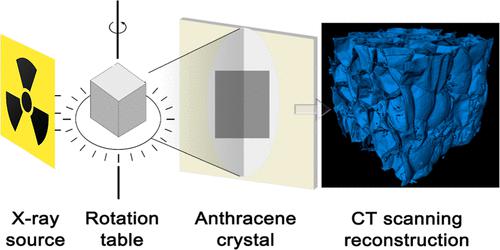
X-ray imaging and computed tomography (CT) technology, as the important non-destructive measurements, can observe internal structures without destroying the detected sample, which are always used in biological diagnosis to detect tumors, pathologies, and bone damages. It is always a challenge to find materials with a low detection limit, a short exposure time, and high resolution to reduce X-ray damage and acquire high-contrast images. Here, we described a low-cost and high-efficient method to prepare centimeter-sized anthracene crystals, which exhibited intense X-ray radioluminescence with a detection limit of ∼0.108 μGy s–1, which is only one-fifth of the dose typically used for X-ray diagnostics. Additionally, the low absorption reduced the damage in radiation and ensured superior cycle performance. X-ray detectors based on anthracene crystals also exhibited an extremely high resolution of 40 lp mm–1. The CT scanning and reconstruction of a foam sample were then achieved, and the detailed internal structure could be clearly observed. These indicated that organic crystals are expecting to be leading candidate low-cost materials for low-dose and highly sensitive X-ray detection and CT scanning.
中文翻译:

用于计算机断层扫描的蒽单晶闪烁体
X射线成像和计算机断层扫描(CT)技术作为重要的无损测量技术,可以在不破坏被检测样本的情况下观察内部结构,在生物诊断中一直用于检测肿瘤、病理和骨损伤。寻找具有低检测限、短曝光时间和高分辨率的材料以减少 X 射线损伤并获取高对比度图像始终是一项挑战。在这里,我们描述了一种低成本、高效的方法来制备厘米大小的蒽晶体,该晶体表现出强烈的 X 射线辐射发光,检测限为 ~0.108 μGy s –1,这只是通常用于 X 射线诊断的剂量的五分之一。此外,低吸收减少了辐射损伤并确保了卓越的循环性能。基于蒽晶体的 X 射线探测器也表现出 40 lp mm –1的极高分辨率。然后实现了泡沫样品的CT扫描和重建,可以清楚地观察到详细的内部结构。这些表明有机晶体有望成为低剂量和高灵敏度 X 射线检测和 CT 扫描的主要候选低成本材料。
更新日期:2022-09-05
中文翻译:

用于计算机断层扫描的蒽单晶闪烁体
X射线成像和计算机断层扫描(CT)技术作为重要的无损测量技术,可以在不破坏被检测样本的情况下观察内部结构,在生物诊断中一直用于检测肿瘤、病理和骨损伤。寻找具有低检测限、短曝光时间和高分辨率的材料以减少 X 射线损伤并获取高对比度图像始终是一项挑战。在这里,我们描述了一种低成本、高效的方法来制备厘米大小的蒽晶体,该晶体表现出强烈的 X 射线辐射发光,检测限为 ~0.108 μGy s –1,这只是通常用于 X 射线诊断的剂量的五分之一。此外,低吸收减少了辐射损伤并确保了卓越的循环性能。基于蒽晶体的 X 射线探测器也表现出 40 lp mm –1的极高分辨率。然后实现了泡沫样品的CT扫描和重建,可以清楚地观察到详细的内部结构。这些表明有机晶体有望成为低剂量和高灵敏度 X 射线检测和 CT 扫描的主要候选低成本材料。


 京公网安备 11010802027423号