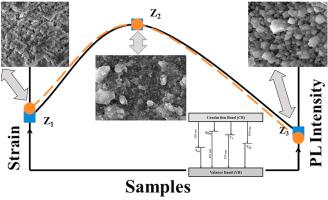
ZnS 和铈 (Ce) 掺杂的 ZnS 的三维 (3-D) 量子结构 (QSs) 是通过化学上负担得起的溶胶-凝胶工艺合成的。探讨了 Ce 掺杂以及由此引起的微应变对结构、形态和光学特性的影响。XRD证实了单相闪锌矿ZnS的形成。对应于最高强度 XRD 峰 (111) 的估计平均晶体尺寸在 1.65-4.65 nm 范围内变化,这与 ZnS 的玻尔半径相当。由于 Ce 和 Zn 的尺寸不匹配,在 ZnS 的基质中发现了微应变和空位。热力学计算验证了由于 Ce 掺杂引起的晶格参数的膨胀和收缩。FTIR 光谱证实了与 Zn 和 S 相关的不同官能团的存在。在 420、461、509和560 nm分别与间隙硫、锌间隙、硫空位和锌空位等缺陷态有关。掺杂 ZnS 在 600 nm 处另一个发射峰的上升是由于Ce 3+离子中的5d → 4f能级跃迁。演化的微应变分布、PL 强度和能带隙变化在掺杂浓度方面彼此相似。显微图像证实了随着掺杂浓度的增加,结构转变为立方体形 ZnS QSs。EDX 和 XPS 支持元素分析以及可用元素(如 Zn、S 和 Ce)的氧化态。
,ZnS 和铈 (Ce) 掺杂的 ZnS 的三维 (3-D) 量子结构 (QSs) 是通过化学上负担得起的溶胶-凝胶工艺合成的。探讨了 Ce 掺杂以及由此引起的微应变对结构、形态和光学特性的影响。XRD证实了单相闪锌矿ZnS的形成。对应于最高强度 XRD 峰 (111) 的估计平均晶体尺寸在 1.65-4.65 nm 范围内变化,这与 ZnS 的玻尔半径相当。由于 Ce 和 Zn 的尺寸不匹配,在 ZnS 的基质中发现了微应变和空位。热力学计算验证了由于 Ce 掺杂引起的晶格参数的膨胀和收缩。FTIR 光谱证实了与 Zn 和 S 相关的不同官能团的存在。在 420、461、509和560 nm分别与间隙硫、锌间隙、硫空位和锌空位等缺陷态有关。掺杂 ZnS 在 600 nm 处另一个发射峰的上升是由于Ce 3+离子中的5d → 4f能级跃迁。演化的微应变分布、PL 强度和能带隙变化在掺杂浓度方面彼此相似。显微图像证实了随着掺杂浓度的增加,结构转变为立方体形 ZnS QSs。EDX 和 XPS 支持元素分析以及可用元素(如 Zn、S 和 Ce)的氧化态。
"点击查看英文标题和摘要"
Interrelation of micro-strain, energy band gap and PL intensity in Ce doped ZnS quantum structures
Three-dimensional (3-D) quantum structures (QSs) of ZnS and Cerium (Ce) doped ZnS were synthesized via chemically affordable sol-gel process. Influence of Ce doping and thus induced micro-strain on the structural, morphological, and optical characteristics was explored. XRD confirmed the formation of single-phase zinc blende ZnS. Estimated average crystal size corresponding to highest intensity XRD peak (111) varied within 1.65–4.65 nm which are comparable with Bohr radius of ZnS. Due to the size mismatch between Ce and Zn, micro-strain and vacancies were found to be developed in host matrix of ZnS. Thermodynamic calculations validated an expansion and contraction in lattice parameter due to Ce doping. FTIR spectra confirmed the presence of different functional groups related to Zn and S. Photoluminescence (PL) emissions observed at 420, 461, 509 and 560 nm are related to the defect states such as interstitial sulfur, zinc interstitial, sulfur vacancies and zinc vacancies respectively. Rise of another emission peak in doped ZnS at 600 nm was due to 5d → 4f energy level transitions in Ce3+ ions. Evolved micro-strain profile, PL intensity and energy band gap variation were analogous to each other with respect to doping concentration. Microscopic images confirmed the structural transformation to cuboidal shaped ZnS QSs with increase in doping concentration. EDX and XPS supported the elemental analysis along with oxidation states of the available elements such as Zn, S and Ce.