Neuroinformatics ( IF 2.7 ) Pub Date : 2022-07-06 , DOI: 10.1007/s12021-022-09588-1 Andres Carrasco 1 , Dorothy E Oorschot 2 , Paolo Barzaghi 3 , Jeffery R Wickens 1
|
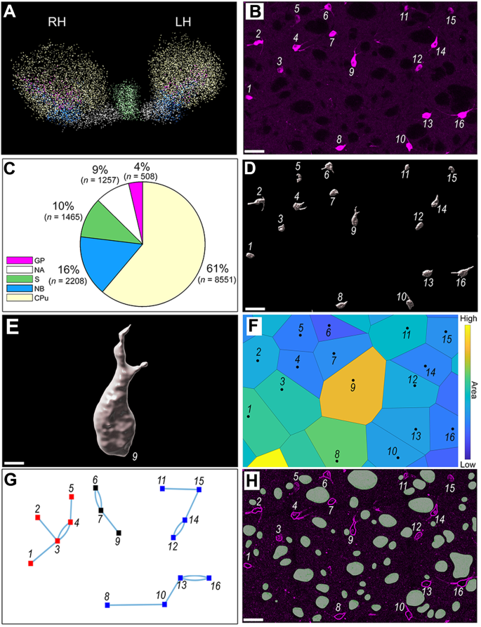
Neuronal networks are regulated by three-dimensional spatial and structural properties. Despite robust evidence of functional implications in the modulation of cognition, little is known about the three-dimensional internal organization of cholinergic networks in the forebrain. Cholinergic networks in the forebrain primarily occur in subcortical nuclei, specifically the septum, nucleus basalis, globus pallidus, nucleus accumbens, and the caudate-putamen. Therefore, the present investigation analyzed the three-dimensional spatial organization of 14,000 cholinergic neurons that expressed choline acetyltransferase (ChAT) in these subcortical nuclei of the mouse forebrain. Point process theory and graph signal processing techniques identified three topological principles of organization. First, cholinergic interneuronal distance is not uniform across brain regions. Specifically, in the septum, globus pallidus, nucleus accumbens, and the caudate-putamen, the cholinergic neurons were clustered compared with a uniform random distribution. In contrast, in the nucleus basalis, the cholinergic neurons had a spatial distribution of greater regularity than a uniform random distribution. Second, a quarter of the caudate-putamen is composed of axonal bundles, yet the spatial distribution of cholinergic neurons remained clustered when axonal bundles were accounted for. However, comparison with an inhomogeneous Poisson distribution showed that the nucleus basalis and caudate-putamen findings could be explained by density gradients in those structures. Third, the number of cholinergic neurons varies as a function of the volume of a specific brain region but cell body volume is constant across regions. The results of the present investigation provide topographic descriptions of cholinergic somata distribution and axonal conduits, and demonstrate spatial differences in cognitive control networks. The study provides a comprehensive digital database of the total population of ChAT-positive neurons in the reported structures, with the x,y,z coordinates of each neuron at micrometer resolution. This information is important for future digital cellular atlases and computational models of the forebrain cholinergic system enabling models based on actual spatial geometry.
中文翻译:
小鼠隔膜、基底核、苍白球、伏隔核和尾壳核胆碱能神经元分布的三维空间分析
神经元网络受三维空间和结构特性的调节。尽管有强有力的证据表明认知调节中的功能影响,但人们对前脑胆碱能网络的三维内部组织知之甚少。前脑中的胆碱能网络主要出现在皮层下核团中,特别是隔膜、基底核、苍白球、伏隔核和尾壳核。因此,本研究分析了 14,000 个在小鼠前脑皮质下核中表达胆碱乙酰转移酶 (ChAT) 的胆碱能神经元的三维空间组织。点过程理论和图形信号处理技术确定了组织的三个拓扑原则。第一的,跨大脑区域的胆碱能神经元间距离不均匀。具体而言,在隔膜、苍白球、伏隔核和尾壳核中,与均匀随机分布相比,胆碱能神经元呈簇状分布。相反,在基底核中,胆碱能神经元的空间分布比均匀随机分布更具规律性。其次,四分之一的尾壳核由轴突束组成,但当考虑到轴突束时,胆碱能神经元的空间分布仍然聚集。然而,与非均匀泊松分布的比较表明,基底核和尾壳核的发现可以用这些结构中的密度梯度来解释。第三,胆碱能神经元的数量随特定大脑区域的体积而变化,但细胞体体积在不同区域是恒定的。本研究的结果提供了胆碱能胞体分布和轴突管道的地形描述,并证明了认知控制网络的空间差异。该研究提供了报告结构中 ChAT 阳性神经元总数的综合数字数据库,其中每个神经元的 x、y、z 坐标都具有微米分辨率。该信息对于未来的数字细胞图谱和前脑胆碱能系统的计算模型非常重要,可支持基于实际空间几何的模型。本研究的结果提供了胆碱能胞体分布和轴突管道的地形描述,并证明了认知控制网络的空间差异。该研究提供了报告结构中 ChAT 阳性神经元总数的综合数字数据库,其中每个神经元的 x、y、z 坐标都具有微米分辨率。该信息对于未来的数字细胞图谱和前脑胆碱能系统的计算模型非常重要,可支持基于实际空间几何的模型。本研究的结果提供了胆碱能胞体分布和轴突管道的地形描述,并证明了认知控制网络的空间差异。该研究提供了报告结构中 ChAT 阳性神经元总数的综合数字数据库,其中每个神经元的 x、y、z 坐标都具有微米分辨率。该信息对于未来的数字细胞图谱和前脑胆碱能系统的计算模型非常重要,可支持基于实际空间几何的模型。每个神经元在微米分辨率下的 z 坐标。该信息对于未来的数字细胞图谱和前脑胆碱能系统的计算模型非常重要,可支持基于实际空间几何的模型。每个神经元在微米分辨率下的 z 坐标。该信息对于未来的数字细胞图谱和前脑胆碱能系统的计算模型非常重要,可支持基于实际空间几何的模型。


 京公网安备 11010802027423号