Our official English website, www.x-mol.net, welcomes your
feedback! (Note: you will need to create a separate account there.)
Ovary structure and symbiotic associates of a ground mealybug, Rhizoecus albidus (Hemiptera, Coccomorpha: Rhizoecidae) and their phylogenetic implications
Journal of Anatomy ( IF 1.8 ) Pub Date : 2022-06-10 , DOI: 10.1111/joa.13712 Teresa Szklarzewicz 1 , Małgorzata Kalandyk-Kołodziejczyk 2 , Anna Michalik 1
Journal of Anatomy ( IF 1.8 ) Pub Date : 2022-06-10 , DOI: 10.1111/joa.13712 Teresa Szklarzewicz 1 , Małgorzata Kalandyk-Kołodziejczyk 2 , Anna Michalik 1
Affiliation

|
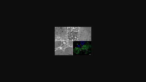
The ovary structure and the organization of its symbiotic system of the ground mealybug, Rhizoecus albidus (Rhizoecidae), were examined by means of microscopic and molecular methods. Each of the paired elongated ovaries of R. albidus is composed of circa one hundred short telotrophic-meroistic ovarioles, which are radially arranged along the distal part of the lateral oviduct. Analysis of serial sections revealed that each ovariole contains four germ cells: three trophocytes (nurse cells) occupying the tropharium and a single oocyte in the vitellarium. The ovaries are accompanied by giant cells termed bacteriocytes which are tightly packed with large pleomorphic bacteria. Their identity as Brownia rhizoecola (Bacteroidetes) was confirmed by means of amplicon sequencing and fluorescence in situ hybridization techniques. Moreover, to our knowledge, this is the first report on the morphology and ultrastructure of the Brownia rhizoecola bacterium. In the bacteriocyte cytoplasm bacteria Brownia co-reside with sporadic rod-shaped smaller bacteria, namely Wolbachia (Proteobacteria: Alphaproteobacteria). Both symbionts are transmitted to the next generation vertically (maternally), that is, via female germline cells. We documented that, at the time when ovarioles contain oocytes at the vitellogenic stage, these symbionts leave the bacteriocytes and move toward the neck region of ovarioles (i.e. the region between tropharium and vitellarium). Next, the bacteria enter the cytoplasm of follicular cells surrounding the basal part of the tropharium, leave them and enter the space between the follicular epithelium and surface of the nutritive cord connecting the tropharium and vitellarium. Finally, they gather in the deep depression of the oolemma at the anterior pole of the oocyte in the form of a ‘symbiont ball’. Our results provide further arguments strongly supporting the validity of the recent changes in the classification of mealybugs, which involved excluding ground mealybugs from the Pseudococcidae family and raising them to the rank of their own family Rhizoecidae.
中文翻译:

地面粉虱的卵巢结构和共生伙伴,Rhizoecus albidus(半翅目,Coccomorpha:Rhizoecidae)及其系统发育意义
通过显微镜和分子方法研究了地面粉虱Rhizoecus albidus (Rhizoecidae)的卵巢结构及其共生系统的组织。 R. albidus的每对细长卵巢均由大约 100 个短端营养性分叶性卵巢组成,这些卵巢沿侧输卵管的远端呈放射状排列。对连续切片的分析显示,每个卵巢管含有四个生殖细胞:三个滋养细胞(护士细胞)占据滋养细胞,一个卵母细胞位于卵黄细胞。卵巢伴随着称为细菌细胞的巨细胞,这些细胞紧密地挤满了大型多形性细菌。通过扩增子测序和荧光原位杂交技术证实了它们的身份为根际布朗氏菌(Bacteroidetes)。此外,据我们所知,这是关于根际布朗氏菌的形态和超微结构的第一份报告。在细菌细胞的细胞质中,布朗尼亚细菌与零星的杆状较小细菌共存,即沃尔巴克氏菌(变形菌门:Alphaproteobacteria)。两种共生体都垂直(母系)传递给下一代,即通过雌性生殖细胞。我们记录到,当卵巢含有处于卵黄发生阶段的卵母细胞时,这些共生体离开细菌细胞并向卵巢的颈部区域(即滋养室和卵黄室之间的区域)移动。接下来,细菌进入滋养层基底部分周围的滤泡细胞的细胞质,离开它们并进入毛囊上皮和连接滋养层和玻璃囊的营养带表面之间的空间。 最后,它们以“共生球”的形式聚集在卵母细胞前极卵膜的凹陷处。我们的结果提供了进一步的论据,有力地支持了近期粉虱分类变化的有效性,其中包括将地面粉虱从拟球虫科中排除,并将它们提升到自己的根螨科的等级。
更新日期:2022-06-10
中文翻译:

地面粉虱的卵巢结构和共生伙伴,Rhizoecus albidus(半翅目,Coccomorpha:Rhizoecidae)及其系统发育意义
通过显微镜和分子方法研究了地面粉虱Rhizoecus albidus (Rhizoecidae)的卵巢结构及其共生系统的组织。 R. albidus的每对细长卵巢均由大约 100 个短端营养性分叶性卵巢组成,这些卵巢沿侧输卵管的远端呈放射状排列。对连续切片的分析显示,每个卵巢管含有四个生殖细胞:三个滋养细胞(护士细胞)占据滋养细胞,一个卵母细胞位于卵黄细胞。卵巢伴随着称为细菌细胞的巨细胞,这些细胞紧密地挤满了大型多形性细菌。通过扩增子测序和荧光原位杂交技术证实了它们的身份为根际布朗氏菌(Bacteroidetes)。此外,据我们所知,这是关于根际布朗氏菌的形态和超微结构的第一份报告。在细菌细胞的细胞质中,布朗尼亚细菌与零星的杆状较小细菌共存,即沃尔巴克氏菌(变形菌门:Alphaproteobacteria)。两种共生体都垂直(母系)传递给下一代,即通过雌性生殖细胞。我们记录到,当卵巢含有处于卵黄发生阶段的卵母细胞时,这些共生体离开细菌细胞并向卵巢的颈部区域(即滋养室和卵黄室之间的区域)移动。接下来,细菌进入滋养层基底部分周围的滤泡细胞的细胞质,离开它们并进入毛囊上皮和连接滋养层和玻璃囊的营养带表面之间的空间。 最后,它们以“共生球”的形式聚集在卵母细胞前极卵膜的凹陷处。我们的结果提供了进一步的论据,有力地支持了近期粉虱分类变化的有效性,其中包括将地面粉虱从拟球虫科中排除,并将它们提升到自己的根螨科的等级。


 京公网安备 11010802027423号