当前位置:
X-MOL 学术
›
J. Mol. Liq.
›
论文详情
Our official English website, www.x-mol.net, welcomes your
feedback! (Note: you will need to create a separate account there.)
Surface fabrication of magnetic core-shell silica nanoparticles with perylene diimide as a fluorescent dye for nucleic acid visualization
Journal of Molecular Liquids ( IF 5.3 ) Pub Date : 2022-05-10 , DOI: 10.1016/j.molliq.2022.119345 Tammar Hussein Ali 1, 2 , Amar Mousa Mandal 3 , Ammar Alhasan 4 , Wim Dehaen 2
Journal of Molecular Liquids ( IF 5.3 ) Pub Date : 2022-05-10 , DOI: 10.1016/j.molliq.2022.119345 Tammar Hussein Ali 1, 2 , Amar Mousa Mandal 3 , Ammar Alhasan 4 , Wim Dehaen 2
Affiliation

|
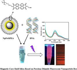
Up to date most traditional fluorescent dyes that serve to detect nucleic acids in a selective manner face growing environmental and economic challenges due to their high toxicity, expensiveness, the long staining times required and the low sensitivities. Further attention went to replace the current widely used ethidium bromide (EB) by more safe and stable dyes but still some of the above drawbacks persist. In this work an innovative fluorescent dye has been designed and synthesized via impregnating perylene diimide into the functionalized surface of magnetic core–shell silica nanoparticles. One of the free hydroxyl groups on the perylene diimide enables the colored magnetic nanoparticles to easily disperse in water and their movement can be controlled with magnetism which allow the separation after their used. These particles are of low toxicity, environmentally friendly and have high sensitivity. Fluorescent properties and molecular interaction were studied. The designed particles show high DNA binding capacity which makes the developed particle promising for DNA extraction, delivery and fluorescent labeling. Theoretical studies confirm that the dye is placed between two DNA base pair similar with pervious observation with EB and the distance was less than 6 Å which is much less than the distance of EB dye interaction (15 Å) as reported before [1] .
中文翻译:

以苝二亚胺为荧光染料制备磁性核壳二氧化硅纳米颗粒用于核酸可视化
迄今为止,大多数用于选择性检测核酸的传统荧光染料由于其高毒性、昂贵、染色时间长和灵敏度低,面临着日益增长的环境和经济挑战。人们进一步关注用更安全稳定的染料取代目前广泛使用的溴化乙锭 (EB),但上述一些缺点仍然存在。在这项工作中,通过将苝二亚胺浸渍到磁性核壳二氧化硅纳米颗粒的功能化表面,设计并合成了一种创新的荧光染料。苝二亚胺上的游离羟基之一使有色磁性纳米颗粒很容易分散在水中,并且它们的运动可以通过磁性控制,从而在使用后进行分离。这些颗粒毒性低、环保且灵敏度高。研究了荧光特性和分子相互作用。设计的颗粒显示出高 DNA 结合载量,这使得开发的颗粒有望用于 DNA 提取、递送和荧光标记。理论研究证实,染料被放置在两个 DNA 碱基对之间,类似于用 EB 进行透视观察,距离小于 6 Å,远小于之前报道的 EB 染料相互作用的距离 (15 Å) [1]。
更新日期:2022-05-10
中文翻译:

以苝二亚胺为荧光染料制备磁性核壳二氧化硅纳米颗粒用于核酸可视化
迄今为止,大多数用于选择性检测核酸的传统荧光染料由于其高毒性、昂贵、染色时间长和灵敏度低,面临着日益增长的环境和经济挑战。人们进一步关注用更安全稳定的染料取代目前广泛使用的溴化乙锭 (EB),但上述一些缺点仍然存在。在这项工作中,通过将苝二亚胺浸渍到磁性核壳二氧化硅纳米颗粒的功能化表面,设计并合成了一种创新的荧光染料。苝二亚胺上的游离羟基之一使有色磁性纳米颗粒很容易分散在水中,并且它们的运动可以通过磁性控制,从而在使用后进行分离。这些颗粒毒性低、环保且灵敏度高。研究了荧光特性和分子相互作用。设计的颗粒显示出高 DNA 结合载量,这使得开发的颗粒有望用于 DNA 提取、递送和荧光标记。理论研究证实,染料被放置在两个 DNA 碱基对之间,类似于用 EB 进行透视观察,距离小于 6 Å,远小于之前报道的 EB 染料相互作用的距离 (15 Å) [1]。


 京公网安备 11010802027423号