Acta Biomaterialia ( IF 9.4 ) Pub Date : 2022-05-10 , DOI: 10.1016/j.actbio.2022.05.010 Zekai Cui 1 , Kai Liao 2 , Shenyang Li 2 , Jianing Gu 1 , Yini Wang 1 , Chengcheng Ding 1 , Yonglong Guo 3 , Hon Fai Chan 4 , Jacey Hongjie Ma 5 , Shibo Tang 6 , Jiansu Chen 7

|
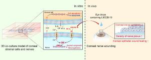
Corneal nerve wounding often causes abnormalities in the cornea and even blindness in severe cases. In this study, we construct a dorsal root ganglion-corneal stromal cell (DRG-CSC, DS) co-culture 3D model to explore the mechanism of corneal nerve regeneration. Firstly, this model consists of DRG collagen grafts sandwiched by orthogonally stacked and orderly arranged CSC-laden plastic compressed collagen. Nerve bundles extend into the entire corneal stroma within 14 days, and they also have orthogonal patterns. This nerve prevents CSCs from apoptosis in the serum withdrawal medium. The conditioned medium (CM) for CSCs in collagen scaffolds contains NT-3, IL-6, and other factors. Among them, NT-3 notably promotes the activation of ERK-CREB in the DRG, leading to the growth of nerve bundles, and IL-6 induces the upregulation of anti-apoptotic genes. Then, LM22B-10, an activator of the NT-3 receptor TrkB/TrkC, can also activate ERK-CREB to enhance nerve growth. After administering LM22B-10 eye drops to regular and diabetic mice with corneal wounding, LM22B-10 significantly improves the healing speed of the corneal epithelium, corneal sensitivity, and corneal nerve density. Overall, the DS co-culture model provides a promising platform and tools for the exploration of corneal physiological and pathological mechanisms, as well as the verification of drug effects in vitro. Meanwhile, we confirm that LM22B-10, as a non-peptide small molecule, has future potential in nerve wound repair.
Statement of significance
The cornea accounts for most of the refractive power of the eye. Corneal nerves play an important role in maintaining corneal homeostasis. Once the corneal nerves are damaged, the corneal epithelium and stroma develop lesions. However, the mechanism of the interaction between corneal nerves and corneal cells is still not fully understood. Here, we construct a corneal stroma-nerve co-culture in vitro model and reveal that NT-3 expressed by stromal cells promotes nerve growth by activating the ERK-CREB pathway in nerves. LM22B-10, an activator of NT-3 receptors, can also induce nerve growth in vitro. Moreover, it is used as eye drops to enhance corneal epithelial wound healing, corneal nerve sensitivity and density of nerve plexus in corneal nerve wounding model in vivo.
中文翻译:

LM22B-10通过体外3D共培养模型和体内角膜损伤模型促进角膜神经再生
角膜神经损伤常导致角膜异常,严重者甚至失明。在这项研究中,我们构建了背根神经节-角膜基质细胞(DRG-CSC,DS)共培养3D模型,以探索角膜神经再生的机制。首先,该模型由正交堆叠和有序排列的载有 CSC 的塑料压缩胶原蛋白夹在 DRG 胶原移植物中。神经束在 14 天内延伸到整个角膜基质,并且它们也具有正交模式。该神经可防止 CSC 在血清戒断培养基中发生凋亡。胶原支架中 CSC 的条件培养基 (CM) 含有 NT-3、IL-6 和其他因子。其中,NT-3显着促进DRG中ERK-CREB的激活,导致神经束生长,IL-6诱导抗凋亡基因上调。然后,LM22B-10 是 NT-3 受体 TrkB/TrkC 的激活剂,也可以激活 ERK-CREB 以增强神经生长。LM22B-10 眼药水对有角膜损伤的常规和糖尿病小鼠给药后,LM22B-10 显着提高了角膜上皮的愈合速度、角膜敏感性和角膜神经密度。总体而言,DS 共培养模型为探索角膜生理和病理机制以及验证药物作用提供了一个有前景的平台和工具。体外。同时,我们确认LM22B-10作为一种非肽类小分子,在神经伤口修复方面具有未来潜力。
重要性声明
角膜占眼睛屈光力的大部分。角膜神经在维持角膜稳态中起重要作用。一旦角膜神经受损,角膜上皮和基质就会出现病变。然而,角膜神经与角膜细胞相互作用的机制仍不完全清楚。在这里,我们构建了一个角膜基质-神经共培养体外模型,并揭示基质细胞表达的 NT-3 通过激活神经中的 ERK-CREB 通路促进神经生长。LM22B-10 是 NT-3 受体的激活剂,也可以在体外诱导神经生长。此外,它还用作眼药水,在体内角膜神经损伤模型中增强角膜上皮伤口愈合、角膜神经敏感性和神经丛密度。.


 京公网安备 11010802027423号