目的
研究磁共振动态磁化率对比灌注加权成像 (DSC-PWI) 和计算机断层扫描灌注 (CTP) 在确定原发性和继发性脑肿瘤的血管分布和渗透性方面的关联和一致性。
材料与方法
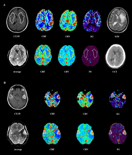
回顾性分析了 97 例高级别胶质瘤、低级别胶质瘤和孤立性脑转移患者的 DSC-PWI 和 CTP 研究。获得归一化脑血流量 (nCBF)、脑血容量 (nCBV)、毛细血管传输常数 (nK2) 和渗透表面积积 (nPS) 值。比较组间变量,测试 DSC-PWI 和 CTP 之间的相关性和一致性。
结果
所有 DSC-PWI 和 CTP 参数在高级别胶质瘤中均高于低级别胶质瘤(p < 0.01 和p < 0.001)。 与低级别胶质瘤相比,转移瘤具有更高的 DSC-PWI nCBV ( p < 0.05)、nCTP-CBF ( p < 0.05)、nCTP-CBV ( p < 0.01) 和 nCTP-PS ( p < 0.0001),并且 nCTP-PS 更高( p < 0.01) 高于高级别神经胶质瘤。DSC-PWI nCBF 和 CTP nCBF 之间(r = 0.79;p < 0.00001)以及 DSC-PWI nCBV 和 CTP nCBV 之间(r = 0.83;p < 0.00001)之间的相关性很强,DSC-PWI nK2 和 CTP nPS 之间的相关性较弱(r = 0.29;p < 0.01)。Bland-Altman 图表明 DSC-PWI nCBF 和 CTP nCBF 之间的一致性很强,DSC-PWI nCBV 和 CTP nCBV 之间的一致性很好,DSC-PWI nK2 和 CTP nPS 之间的一致性较差。
结论
DSC-PWI 和 CTP CBF 和 CBV 图在肿瘤血管分布的评估中具有可比性和可互换性,不像 DSC-PWI K2 和 CTP PS 图在肿瘤通透性分析中更加不一致。当 MRI 不可用或患者不能耐受时,CTP 可能是量化肿瘤新血管生成的替代方法。
 "点击查看英文标题和摘要"
Assessment of brain tumors by magnetic resonance dynamic susceptibility contrast perfusion-weighted imaging and computed tomography perfusion: a comparison study
Purpose
To investigate the association and agreement between magnetic resonance dynamic susceptibility contrast perfusion-weighted imaging (DSC-PWI) and computed tomography perfusion (CTP) in determining vascularity and permeability of primary and secondary brain tumors.
Material and methods
DSC-PWI and CTP studies from 97 patients with high-grade glioma, low-grade glioma and solitary brain metastasis were retrospectively reviewed. Normalized cerebral blood flow (nCBF), cerebral blood volume (nCBV), capillary transfer constant (nK2) and permeability surface area product (nPS) values were obtained. Variables among groups were compared, and correlation and agreement between DSC-PWI and CTP were tested.
Results
All DSC-PWI and CTP parameters were higher in high-grade than in low-grade gliomas (p < 0.01 and p < 0.001). Metastases had greater DSC-PWI nCBV (p < 0.05), nCTP-CBF (p < 0.05), nCTP-CBV (p < 0.01) and nCTP-PS (p < 0.0001) than low-grade gliomas and more elevated nCTP-PS (p < 0.01) than high-grade gliomas. The correlation was strong between DSC-PWI nCBF and CTP nCBF (r = 0.79; p < 0.00001) and between DSC-PWI nCBV and CTP nCBV (r = 0.83; p < 0.00001), weaker between DSC-PWI nK2 and CTP nPS (r = 0.29; p < 0.01). Bland–Altman plots indicated that the agreement was strong between DSC-PWI nCBF and CTP nCBF, good between DSC-PWI nCBV and CTP nCBV and poorer between DSC-PWI nK2 and CTP nPS.
Conclusion
DSC-PWI and CTP CBF and CBV maps were comparable and interchangeable in the assessment of tumor vascularity, unlike DSC-PWI K2 and CTP PS maps that were more discordant in the analysis of tumor permeability. CTP could be an alternative method to quantify tumor neoangiogenesis when MRI is not available or when the patient does not tolerate it.


 京公网安备 11010802027423号