Brazilian Journal of Physics ( IF 1.5 ) Pub Date : 2022-04-18 , DOI: 10.1007/s13538-022-01102-x
R. Dawn 1 , M. Zzaman 1, 2 , A. Kumari 1 , V. R. Singh 1 , R. Shahid 2 , F. Faizal 3, 4 , C. Panatarani 3, 4 , I. M. Joni 3, 4 , C. Kiran 5 , V. K. Verma 6 , S. K. Sahoo 7 , K. Amemiya 8
|
Abstract
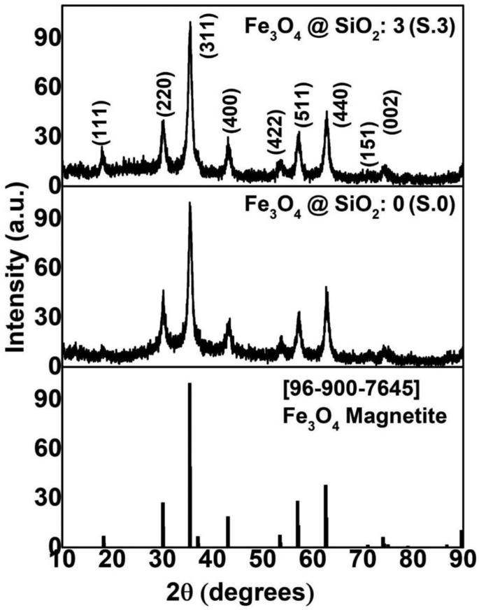
Magnetite (Fe3O4) nanoparticles (NPs) and SiO2-coated Fe3O4 nanoparticles have successfully been synthesized using co-precipitation and modified Stöber methods, respectively. The samples were characterized using X-ray diffraction (XRD), Fourier transform infrared (FTIR) spectroscopy, high-resolution transmission electron microscopy (HRTEM), vibrating sample magnetometer (VSM) techniques, X-ray absorption spectroscopy (XAS), and X-ray magnetic circular dichroism (XMCD). XRD and FTIR data confirmed the structural configuration of a single-phase Fe3O4 and the successful formation of SiO2-coated Fe3O4 NPs. XRD also confirmed that we have succeeded to synthesize nano-meter size of Fe3O4 NPs. HRTEM images showed the increasing thickness of SiO2-coated Fe3O4 with the addition of the Tetraethyl Orthosilicate (TEOS). Room temperature VSM analysis showed the magnetic behaviour of Fe3O4 and its variations that occurred after SiO2 coating. The magnetic behaviour is further authenticated by XAS spectra analysis which cleared about the existence of SiO2 shells that have transformed the crystal as well as the local structures of the magnetite NPs. We have performed XMCD measurements, which is a powerful element-specific technique to find out the origin of magnetization in SiO2-coated Fe3O4 NPs, that verified a decrease in magnetization with increasing thickness of the SiO2 coating.
Graphical Abstract
Magnetite (Fe3O4) nanoparticles (NPs) and SiO2-coated Fe3O4 nanoparticles have successfully been synthesized using co-precipitation and modified Stöber methods, respectively. The samples were characterized using X-ray diffraction (XRD), Fourier transform infrared (FTIR) spectroscopy, high-resolution transmission electron microscopy (HRTEM), vibrating sample magnetometer (VSM) techniques, X-ray absorption spectroscopy (XAS), and X-ray magnetic circular dichroism (XMCD). XRD and FTIR data confirmed the structural configuration of a single-phase Fe3O4 and the successful formation of SiO2-coated Fe3O4 NPs. XRD also confirmed that we have succeeded to synthesize nano-meter size of Fe3O4 NPs. HRTEM images showed the increasing thickness of SiO2-coated Fe3O4 with the addition of the Tetraethyl Orthosilicate (TEOS). Room temperature VSM analysis showed the magnetic behaviour of Fe3O4 and its variations that occurred after SiO2 coating. The magnetic behaviour is further authenticated by XAS spectra analysis which cleared about the existence of SiO2 shells that have transformed the crystal as well as the local structures of the magnetite NPs. We have performed XMCD measurements, which is a powerful element-specific technique to find out the origin of magnetization in SiO2-coated Fe3O4 NPs, that verified a decrease in magnetization with increasing thickness of the SiO2 coating.
中文翻译:
软X射线磁圆二色性揭示二氧化硅包覆Fe3O4纳米颗粒的磁化起源
摘要
磁铁矿 (Fe 3 O 4 ) 纳米粒子 (NPs) 和 SiO 2包覆的 Fe 3 O 4纳米粒子已分别使用共沉淀法和改进的 Stöber 方法成功合成。使用 X 射线衍射 (XRD)、傅里叶变换红外 (FTIR) 光谱、高分辨率透射电子显微镜 (HRTEM)、振动样品磁力计 (VSM) 技术、X 射线吸收光谱 (XAS) 和 X射线磁圆二色性(XMCD)。XRD和FTIR数据证实了单相Fe 3 O 4的结构构型和SiO 2包覆的Fe 3 O的成功形成4个NP。XRD还证实我们已经成功合成了纳米尺寸的Fe 3 O 4 NPs。HRTEM 图像显示随着原硅酸四乙酯 (TEOS) 的添加, SiO 2包覆的 Fe 3 O 4的厚度增加。室温 VSM 分析显示了 Fe 3 O 4的磁行为及其在 SiO 2涂层后发生的变化。XAS 光谱分析进一步证实了磁性行为,该分析明确了 SiO 2的存在改变晶体的壳以及磁铁矿NP的局部结构。我们进行了 XMCD 测量,这是一种强大的元素特异性技术,可以找出 SiO 2 涂层的 Fe 3 O 4 NPs 中磁化的起源,这证实了磁化强度随着 SiO 2涂层厚度的增加而降低。
图形概要
磁铁矿 (Fe 3 O 4 ) 纳米粒子 (NPs) 和 SiO 2包覆的 Fe 3 O 4纳米粒子已分别使用共沉淀法和改进的 Stöber 方法成功合成。使用 X 射线衍射 (XRD)、傅里叶变换红外 (FTIR) 光谱、高分辨率透射电子显微镜 (HRTEM)、振动样品磁力计 (VSM) 技术、X 射线吸收光谱 (XAS) 和 X射线磁圆二色性(XMCD)。XRD和FTIR数据证实了单相Fe 3 O 4的结构构型和SiO 2包覆的Fe 3 O的成功形成4个NP。XRD还证实我们已经成功合成了纳米尺寸的Fe 3 O 4 NPs。HRTEM 图像显示随着原硅酸四乙酯 (TEOS) 的添加, SiO 2包覆的 Fe 3 O 4的厚度增加。室温 VSM 分析显示了 Fe 3 O 4的磁行为及其在 SiO 2涂层后发生的变化。XAS 光谱分析进一步证实了磁性行为,该分析明确了 SiO 2的存在改变晶体的壳以及磁铁矿NP的局部结构。我们进行了 XMCD 测量,这是一种强大的元素特异性技术,可以找出 SiO 2 涂层的 Fe 3 O 4 NPs 中磁化的起源,这证实了磁化强度随着 SiO 2涂层厚度的增加而降低。


 京公网安备 11010802027423号