Dental Materials ( IF 4.6 ) Pub Date : 2021-12-23 , DOI: 10.1016/j.dental.2021.12.135 Jinmeng Li 1 , Abeer Ahmed 2 , Tanika Degrande 3 , Jérémie De Baerdemaeker 3 , Abdulaziz Al-Rasheed 2 , Jeroen Jjp van den Beucken 1 , John A Jansen 1 , Hamdan S Alghamdi 4 , X Frank Walboomers 1

|
Objectives
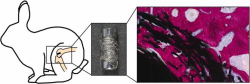
This study was aimed to comparatively evaluate new bone formation into the pores of a flexible titanium fiber mesh (TFM) applied on the surface of implant.
Methods
Twenty-eight custom made cylindrical titanium implants (4 ×10 mm) with and without a layer of two different types of TFM (fiber diameter of 22 µm and 50 µm, volumetric porosity ~70%) were manufactured and installed bilaterally in the femoral condyles of 14 rabbits. The elastic modulus for these two TFM types was ~20 GPa and ~5 GPa respectively, whereas the solid titanium was ~110 GPa. The implants (Control, TFM-22, TFM-50) were retrieved after 14 weeks of healing and prepared for histological assessment. The percentage of the bone area (BA%), the bone-to-implant contact (BIC%) and amount were determined.
Results
Newly formed bone into mesh porosity was observed for all three types of implants. Histomorphometric analyses revealed significantly higher (~2.5 fold) BA% values for TFM-22 implants (30.9 ± 9.5%) compared to Control implants (12.7 ± 6.0%), whereas BA% for TMF-50 did not significantly differ compared with Control implants. Furthermore, both TFM-22 and TFM-50 implants showed significantly higher BIC% values (64.9 ± 14.0%, ~2.5 fold; 47.1 ± 14.1%, ~2 fold) compared to Control (23.6 ± 17.4%). Finally, TFM-22 implants showed more and thicker trabeculae in the peri-implant region.
Significance
This in vivo study demonstrated that implants with a flexible coating of TFM improve bone formation within the inter-fiber space and the peri-implant region.
中文翻译:

兔股骨髁模型中钛纤维网状涂层植入物的组织学评价
目标
本研究旨在比较评估新骨在植入物表面应用的柔性钛纤维网 (TFM) 孔隙中的形成。
方法
制造了 28 个定制的圆柱形钛植入物 (4 × 10 mm),带有和不带有两种不同类型的 TFM(纤维直径为 22 µm 和 50 µm,体积孔隙率 ~70%)的层,并在双侧股骨髁中安装14 只兔子。这两种 TFM 类型的弹性模量分别为 ~20 GPa 和 ~5 GPa,而固态钛为 ~110 GPa。在愈合 14 周后取出植入物(对照、TFM-22、TFM-50)并准备进行组织学评估。确定骨面积百分比 (BA%)、骨与种植体接触 (BIC%) 和数量。
结果
对于所有三种类型的植入物,都观察到新形成的骨成网孔。组织形态学分析显示,与对照植入物(12.7 ± 6.0%)相比,TFM-22 植入物的 BA% 值(30.9 ± 9.5%)显着更高(~2.5 倍),而 TMF-50 的 BA% 与对照植入物相比没有显着差异. 此外,与对照(23.6 ± 17.4%)相比,TFM-22 和 TFM-50 植入物的 BIC% 值(64.9 ± 14.0%,~2.5 倍;47.1 ± 14.1%,~2 倍)显着更高。最后,TFM-22 种植体在种植体周围区域显示出更多和更厚的小梁。
意义
这项体内研究表明,具有柔性 TFM 涂层的植入物可以改善纤维间空间和植入物周围区域的骨形成。


 京公网安备 11010802027423号