Our official English website, www.x-mol.net, welcomes your
feedback! (Note: you will need to create a separate account there.)
Histomorphometry of mast cells in the convexity of human intracranial dura mater
Journal of Anatomy ( IF 1.8 ) Pub Date : 2021-11-23 , DOI: 10.1111/joa.13585 Emanuela P Rosas 1 , Silvania T Paz 2 , Raisa F Costa 3 , Amanda A da Silva 4 , Romildo L da Silva 5 , Ana P F da Silva 1 , Sabrina R S da Silva 6 , Paloma L de Medeiros 2 , Manuela F L de Freitas 7 , Marcelo M Valença 1, 3, 4
Journal of Anatomy ( IF 1.8 ) Pub Date : 2021-11-23 , DOI: 10.1111/joa.13585 Emanuela P Rosas 1 , Silvania T Paz 2 , Raisa F Costa 3 , Amanda A da Silva 4 , Romildo L da Silva 5 , Ana P F da Silva 1 , Sabrina R S da Silva 6 , Paloma L de Medeiros 2 , Manuela F L de Freitas 7 , Marcelo M Valença 1, 3, 4
Affiliation
|
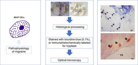
Mast cells, known as pro-inflammatory effector cells, are immunocytes present in the meninges and may be involved in the pathophysiology of migraine. This study aims to evaluate the histomorphometric parameters of mast cells located in the convexity of the human intracranial dura mater. For this, samples of intracranial dura mater from eight human fresh cadavers were collected between 8- and 24-h post-mortem. The whole samples were fixed and, subsequently, two fragments of 1.5 cm² each were cut from four different areas of the dura mater convexity, containing a segment of the middle meningeal artery, totaling 64 fragments. After histological processing, the fragments were submitted to microtomy (5 and 10 µm), stained with toluidine blue (0.1%), or immunohistochemically labeled for tryptase, and analyzed using optical microscopy. The following histomorphometric parameters were evaluated: distance from mast cells to vessels, the density of mast cells, and percentage of mast cells with degranulation. Histomorphometric analyzes showed a higher density of mast cells in the vicinity of blood vessels (arterial and venous), with distances around 0–150 µm. A greater number of mast cells was detected near venous vessels in the periosteal layer (17.0 ± 10.1 cells/mm²) than in the meningeal layer (14.1 ± 7.0 cells/mm²) (p < 0.05). Mast cells from the region close to the superior sagittal sinus were found in greater quantity close to the venous vessels (16.7 ± 10.1 cells/mm²) than to the arterial vessels (11.2 ± 7.5 cells/mm²) (p < 0.05). In short, in the convexity of the human intracranial dura mater, mast cells are located close to blood vessels, with a greater number of cells next to the venous vessels of the periosteal layer and in the proximal region of the superior sagittal sinus.
中文翻译:

人颅内硬脑膜凸面肥大细胞的组织形态计量学
肥大细胞,称为促炎效应细胞,是存在于脑膜中的免疫细胞,可能参与偏头痛的病理生理学。本研究旨在评估位于人颅内硬脑膜凸面的肥大细胞的组织形态学参数。为此,我们在死后 8 至 24 小时内采集了 8 具新鲜人类尸体的颅内硬脑膜样本。将整个样本固定,随后从硬脑膜凸面的四个不同区域切下两个各1.5cm²的碎片,其中包含一段脑膜中动脉,总共64个碎片。组织学处理后,将片段进行切片(5 和 10 µm),用甲苯胺蓝(0.1%)染色,或对类胰蛋白酶进行免疫组织化学标记,并使用光学显微镜进行分析。评估以下组织形态学参数:肥大细胞到血管的距离、肥大细胞的密度以及脱颗粒的肥大细胞的百分比。组织形态计量学分析显示,血管(动脉和静脉)附近的肥大细胞密度较高,距离约为 0-150 µm。在骨膜层静脉血管附近检测到的肥大细胞数量 (17.0 ± 10.1 个细胞/mm2) 多于脑膜层 (14.1 ± 7.0 个细胞/mm2) ( p < 0.05)。靠近上矢状窦区域的肥大细胞靠近静脉血管的数量(16.7 ± 10.1 个细胞/mm²)多于靠近动脉血管的数量(11.2 ± 7.5 个细胞/mm²)( p < 0.05)。 总之,在人颅内硬脑膜的凸面,肥大细胞靠近血管,其中靠近骨膜层静脉血管和上矢状窦近端区域的细胞数量较多。
更新日期:2021-11-23
中文翻译:
人颅内硬脑膜凸面肥大细胞的组织形态计量学
肥大细胞,称为促炎效应细胞,是存在于脑膜中的免疫细胞,可能参与偏头痛的病理生理学。本研究旨在评估位于人颅内硬脑膜凸面的肥大细胞的组织形态学参数。为此,我们在死后 8 至 24 小时内采集了 8 具新鲜人类尸体的颅内硬脑膜样本。将整个样本固定,随后从硬脑膜凸面的四个不同区域切下两个各1.5cm²的碎片,其中包含一段脑膜中动脉,总共64个碎片。组织学处理后,将片段进行切片(5 和 10 µm),用甲苯胺蓝(0.1%)染色,或对类胰蛋白酶进行免疫组织化学标记,并使用光学显微镜进行分析。评估以下组织形态学参数:肥大细胞到血管的距离、肥大细胞的密度以及脱颗粒的肥大细胞的百分比。组织形态计量学分析显示,血管(动脉和静脉)附近的肥大细胞密度较高,距离约为 0-150 µm。在骨膜层静脉血管附近检测到的肥大细胞数量 (17.0 ± 10.1 个细胞/mm2) 多于脑膜层 (14.1 ± 7.0 个细胞/mm2) ( p < 0.05)。靠近上矢状窦区域的肥大细胞靠近静脉血管的数量(16.7 ± 10.1 个细胞/mm²)多于靠近动脉血管的数量(11.2 ± 7.5 个细胞/mm²)( p < 0.05)。 总之,在人颅内硬脑膜的凸面,肥大细胞靠近血管,其中靠近骨膜层静脉血管和上矢状窦近端区域的细胞数量较多。


 京公网安备 11010802027423号