当前位置:
X-MOL 学术
›
Mol. Ther.
›
论文详情
Our official English website, www.x-mol.net, welcomes your
feedback! (Note: you will need to create a separate account there.)
Spatial distribution and functional analysis define the action pathway of Tim-3/Tim-3 ligands in tumor development
Molecular Therapy ( IF 12.1 ) Pub Date : 2021-11-19 , DOI: 10.1016/j.ymthe.2021.11.015
Tixiao Wang 1 , Jie Zhang 1 , Na Li 1 , Mengzhen Li 1 , Shuaiya Ma 1 , Siyu Tan 1 , Xiaowei Guo 1 , Zehua Wang 1 , Zhuanchang Wu 1 , Lifen Gao 1 , Chunhong Ma 2 , Xiaohong Liang 2
Molecular Therapy ( IF 12.1 ) Pub Date : 2021-11-19 , DOI: 10.1016/j.ymthe.2021.11.015
Tixiao Wang 1 , Jie Zhang 1 , Na Li 1 , Mengzhen Li 1 , Shuaiya Ma 1 , Siyu Tan 1 , Xiaowei Guo 1 , Zehua Wang 1 , Zhuanchang Wu 1 , Lifen Gao 1 , Chunhong Ma 2 , Xiaohong Liang 2
Affiliation
|
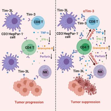
The spatial organization of immune cells within the tumor microenvironment (TME) largely determines the anti-tumor immunity and also highly predicts tumor progression and therapeutic response. Tim-3 is a well-accepted immune checkpoint and plays multifaceted immunoregulatory roles via interaction with distinct Tim-3 ligands (Tim-3L), showing great potential as an immunotherapy target. However, the cell sociology mediated by Tim-3/Tim-3L and their contribution to tumor development remains elusive. Here, we analyzed the spatial distribution of Tim-3/Tim-3L in TME using multiplex fluorescence staining and revealed that despite the increased Tim-3 expression in various tumor-infiltrated lymphocytes, Tim-3CD4 cells were more accumulated in parenchymal/tumor region compared with stromal region and exhibited more close association with patient survival. Strikingly, CD4 T cells surrounding Tim-3L cells expressed higher Tim-3 than other cells in cancerous tissues. studies confirmed that depletion of CD4 T cells completely abrogated the inhibition of tumor growth and metastasis, as well as the functional improvement of CD8 T and NK, mediated by Tim-3 blockade, which was further validated in peripheral lymphocytes from patients with hepatocellular carcinoma. In conclusion, our findings unravel the importance of CD4 T cells in Tim-3/Tim-3L-mediated immunosuppression and provide new thoughts for Tim-3 targeted cancer immunotherapy.
中文翻译:

空间分布和功能分析定义了 Tim-3/Tim-3 配体在肿瘤发展中的作用途径
肿瘤微环境(TME)内免疫细胞的空间组织在很大程度上决定了抗肿瘤免疫力,并且高度预测肿瘤进展和治疗反应。 Tim-3 是一种广为接受的免疫检查点,通过与不同的 Tim-3 配体 (Tim-3L) 相互作用发挥多方面的免疫调节作用,显示出作为免疫治疗靶点的巨大潜力。然而,Tim-3/Tim-3L 介导的细胞社会学及其对肿瘤发展的贡献仍然难以捉摸。在这里,我们使用多重荧光染色分析了 TME 中 Tim-3/Tim-3L 的空间分布,发现尽管各种肿瘤浸润淋巴细胞中 Tim-3 表达增加,但 Tim-3CD4 细胞在实质/肿瘤区域聚集更多与间质区域相比,与患者生存具有更密切的相关性。引人注目的是,Tim-3L 细胞周围的 CD4 T 细胞比癌组织中的其他细胞表达更高的 Tim-3。研究证实,CD4 T 细胞的耗竭完全消除了对肿瘤生长和转移的抑制,以及由 Tim-3 阻断介导的 CD8 T 和 NK 功能的改善,这在肝细胞癌患者的外周淋巴细胞中得到了进一步验证。总之,我们的研究结果揭示了 CD4 T 细胞在 Tim-3/Tim-3L 介导的免疫抑制中的重要性,并为 Tim-3 靶向癌症免疫治疗提供了新思路。
更新日期:2021-11-19
中文翻译:
空间分布和功能分析定义了 Tim-3/Tim-3 配体在肿瘤发展中的作用途径
肿瘤微环境(TME)内免疫细胞的空间组织在很大程度上决定了抗肿瘤免疫力,并且高度预测肿瘤进展和治疗反应。 Tim-3 是一种广为接受的免疫检查点,通过与不同的 Tim-3 配体 (Tim-3L) 相互作用发挥多方面的免疫调节作用,显示出作为免疫治疗靶点的巨大潜力。然而,Tim-3/Tim-3L 介导的细胞社会学及其对肿瘤发展的贡献仍然难以捉摸。在这里,我们使用多重荧光染色分析了 TME 中 Tim-3/Tim-3L 的空间分布,发现尽管各种肿瘤浸润淋巴细胞中 Tim-3 表达增加,但 Tim-3CD4 细胞在实质/肿瘤区域聚集更多与间质区域相比,与患者生存具有更密切的相关性。引人注目的是,Tim-3L 细胞周围的 CD4 T 细胞比癌组织中的其他细胞表达更高的 Tim-3。研究证实,CD4 T 细胞的耗竭完全消除了对肿瘤生长和转移的抑制,以及由 Tim-3 阻断介导的 CD8 T 和 NK 功能的改善,这在肝细胞癌患者的外周淋巴细胞中得到了进一步验证。总之,我们的研究结果揭示了 CD4 T 细胞在 Tim-3/Tim-3L 介导的免疫抑制中的重要性,并为 Tim-3 靶向癌症免疫治疗提供了新思路。


 京公网安备 11010802027423号