当前位置:
X-MOL 学术
›
Magn. Reson. Imaging
›
论文详情
Our official English website, www.x-mol.net, welcomes your feedback! (Note: you will need to create a separate account there.)
T2 Relaxometry Evidence of Microstructural Changes in Diffusely Abnormal White Matter in Relapsing–Remitting Multiple Sclerosis and Clinically Isolated Syndrome: Impact on Visuomotor Performance
Journal of Magnetic Resonance Imaging ( IF 3.3 ) Pub Date : 2021-09-14 , DOI: 10.1002/jmri.27233 Efrosini Papadaki , Vasileios Mastorodemos , Theodora Panou , Styliani Pouli , Eirini Spyridaki , Eleftherios Kavroulakis , Georgios Kalaitzakis , Thomas G. Maris , Panagiotis Simos
Journal of Magnetic Resonance Imaging ( IF 3.3 ) Pub Date : 2021-09-14 , DOI: 10.1002/jmri.27233 Efrosini Papadaki , Vasileios Mastorodemos , Theodora Panou , Styliani Pouli , Eirini Spyridaki , Eleftherios Kavroulakis , Georgios Kalaitzakis , Thomas G. Maris , Panagiotis Simos
|
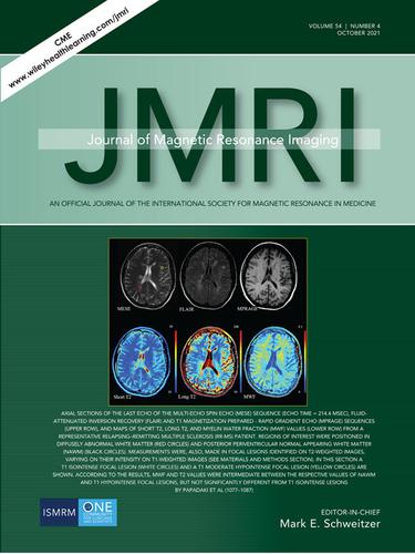
Axial sections of the last echo of the multi-echo spin echo (MESE) sequence (echo time = 214.4 MSEC), fluidattenuated inversion recovery (Flair) and T1 magnetization prepared - rapid gradient echo (MPRAGE) sequences (Upper Row), and maps of short t2, long t2, and myelin water fraction (MWF) values (Lower Row) from a representative relapsing–remitting multiple sclerosis (RR-MS) patient. Regions of interest were positioned in diffusely abnormal white matter (Red Circles) and posterior periventricular normal appearing white matter (NAWM) (Black Circles). Measurements were, also, made in focal lesions identified on T2-weighted images, varying on their intensity on T1-weighted images (see materials and methods section). In this section A T1 isointense focal lesion (White Circles) and a T1 moderate hypointense focal lesion (Yellow Circles) are shown. According to the results, MWF and T2 values were intermediate between the respective values of NAWM and T1 hypointense focal lesions, but not significantly different from T1 isointense lesions by papadaki et al (1077–1087)

中文翻译:

复发缓解型多发性硬化症和临床孤立综合征中弥漫性异常白质微结构变化的 T2 松弛测量证据:对视觉运动表现的影响
多回波自旋回波 (MESE) 序列(回波时间 = 214.4 MSEC)、流体衰减反转恢复 (Flair) 和 T1 磁化准备的最后回波的轴向截面 - 快速梯度回波 (MPRAGE) 序列(上排)和地图来自代表性复发缓解型多发性硬化症 (RR-MS) 患者的短 t2、长 t2 和髓鞘水分数 (MWF) 值(下排)。感兴趣的区域位于弥漫性异常白质(红圈)和后脑室周围正常出现的白质(NAWM)(黑圈)中。测量也是在 T2 加权图像上识别的局灶性病变中进行的,它们在 T1 加权图像上的强度不同(参见材料和方法部分)。在本节中,显示了 T1 等信号局灶性病变(白色圆圈)和 T1 中度低信号局灶性病变(黄色圆圈)。
更新日期:2021-09-14
中文翻译:
复发缓解型多发性硬化症和临床孤立综合征中弥漫性异常白质微结构变化的 T2 松弛测量证据:对视觉运动表现的影响
多回波自旋回波 (MESE) 序列(回波时间 = 214.4 MSEC)、流体衰减反转恢复 (Flair) 和 T1 磁化准备的最后回波的轴向截面 - 快速梯度回波 (MPRAGE) 序列(上排)和地图来自代表性复发缓解型多发性硬化症 (RR-MS) 患者的短 t2、长 t2 和髓鞘水分数 (MWF) 值(下排)。感兴趣的区域位于弥漫性异常白质(红圈)和后脑室周围正常出现的白质(NAWM)(黑圈)中。测量也是在 T2 加权图像上识别的局灶性病变中进行的,它们在 T1 加权图像上的强度不同(参见材料和方法部分)。在本节中,显示了 T1 等信号局灶性病变(白色圆圈)和 T1 中度低信号局灶性病变(黄色圆圈)。


 京公网安备 11010802027423号