当前位置:
X-MOL 学术
›
J. Orthop. Res.
›
论文详情
Our official English website, www.x-mol.net, welcomes your
feedback! (Note: you will need to create a separate account there.)
MRI- and CT-based metrics for the quantification of arthroscopic bone resections in femoroacetabular impingement syndrome
Journal of Orthopaedic Research ( IF 2.1 ) Pub Date : 2021-06-30 , DOI: 10.1002/jor.25139 Martina Guidetti 1 , Philip Malloy 1, 2 , Thomas D Alter 1 , Alexander C Newhouse 1 , Alejandro A Espinoza Orías 1 , Nozomu Inoue 1 , Shane J Nho 1
Journal of Orthopaedic Research ( IF 2.1 ) Pub Date : 2021-06-30 , DOI: 10.1002/jor.25139 Martina Guidetti 1 , Philip Malloy 1, 2 , Thomas D Alter 1 , Alexander C Newhouse 1 , Alejandro A Espinoza Orías 1 , Nozomu Inoue 1 , Shane J Nho 1
Affiliation

|
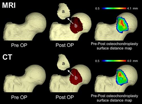
The purpose of this in vitro study was to quantify the bone resected from the proximal femur during hip arthroscopy using metrics generated from magnetic resonance imaging (MRI) and computed tomography (CT) reconstructed threedimensional (3D) bone models. Seven cadaveric hemipelvises underwent both a 1.5 T MRI and CT scan before and following an arthroscopic proximal femoral osteochondroplasty. The images from MRI and CT were segmented to generate 3D proximal femoral surface models. A validated 3D-3D registration method was used to compare surface-to-surface distances between the 3D models before and following surgery. The new metrics of maximum height, mean height, surface area and volume, were computed to quantify bone resected during osteochondroplasty. Stability of the metrics across imaging modalities was established through paired sample t-tests and bivariate correlation. Bivariate correlation analyses indicated strong correlations between all metrics (r = 0.728–0.878) computed from MRI and CT derived models. There were no differences in the MRI- and CT-based metrics used to quantify bone resected during femoral osteochondroplasty. Preoperative and postoperative MRI and CT derived 3D bone models can be used to quantify bone resected during femoral osteochondroplasty, without significant differences between the imaging modalities.
中文翻译:

基于 MRI 和 CT 的指标用于量化股骨髋臼撞击综合征的关节镜下骨切除术
这项体外研究的目的是使用从磁共振成像 (MRI) 和计算机断层扫描 (CT) 重建的三维 (3D) 骨骼模型生成的指标来量化髋关节镜检查期间从股骨近端切除的骨骼。七具尸体半骨盆在关节镜下股骨近端骨软骨成形术之前和之后都接受了 1.5 T MRI 和 CT 扫描。对来自 MRI 和 CT 的图像进行分割以生成 3D 股骨近端表面模型。使用经过验证的 3D-3D 配准方法来比较手术前后 3D 模型之间的表面到表面距离。计算最大高度、平均高度、表面积和体积的新指标以量化骨软骨成形术期间切除的骨。通过配对样本 t 检验和双变量相关性建立跨成像模式的指标的稳定性。双变量相关分析表明所有指标之间存在很强的相关性(r = 0.728–0.878)从 MRI 和 CT 衍生模型计算得出。用于量化股骨骨软骨成形术期间切除的骨的基于 MRI 和 CT 的指标没有差异。术前和术后 MRI 和 CT 衍生的 3D 骨模型可用于量化股骨软骨成形术期间切除的骨,成像方式之间没有显着差异。
更新日期:2021-06-30
中文翻译:

基于 MRI 和 CT 的指标用于量化股骨髋臼撞击综合征的关节镜下骨切除术
这项体外研究的目的是使用从磁共振成像 (MRI) 和计算机断层扫描 (CT) 重建的三维 (3D) 骨骼模型生成的指标来量化髋关节镜检查期间从股骨近端切除的骨骼。七具尸体半骨盆在关节镜下股骨近端骨软骨成形术之前和之后都接受了 1.5 T MRI 和 CT 扫描。对来自 MRI 和 CT 的图像进行分割以生成 3D 股骨近端表面模型。使用经过验证的 3D-3D 配准方法来比较手术前后 3D 模型之间的表面到表面距离。计算最大高度、平均高度、表面积和体积的新指标以量化骨软骨成形术期间切除的骨。通过配对样本 t 检验和双变量相关性建立跨成像模式的指标的稳定性。双变量相关分析表明所有指标之间存在很强的相关性(r = 0.728–0.878)从 MRI 和 CT 衍生模型计算得出。用于量化股骨骨软骨成形术期间切除的骨的基于 MRI 和 CT 的指标没有差异。术前和术后 MRI 和 CT 衍生的 3D 骨模型可用于量化股骨软骨成形术期间切除的骨,成像方式之间没有显着差异。


 京公网安备 11010802027423号