当前位置:
X-MOL 学术
›
Anal. Chem.
›
论文详情
Our official English website, www.x-mol.net, welcomes your
feedback! (Note: you will need to create a separate account there.)
Multiplexed Ion Beam Imaging Readout of Single-Cell Immunoblotting
Analytical Chemistry ( IF 6.7 ) Pub Date : 2021-06-09 , DOI: 10.1021/acs.analchem.1c01050
Gabriela Lomeli , Marc Bosse 1 , Sean C Bendall 1 , Michael Angelo 1 , Amy E Herr 2
Analytical Chemistry ( IF 6.7 ) Pub Date : 2021-06-09 , DOI: 10.1021/acs.analchem.1c01050
Gabriela Lomeli , Marc Bosse 1 , Sean C Bendall 1 , Michael Angelo 1 , Amy E Herr 2
Affiliation
|
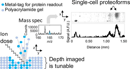
Improvements in single-cell protein analysis are required to study the cell-to-cell variation inherent to diseases, including cancer. Single-cell immunoblotting (scIB) offers proteoform detection specificity, but often relies on fluorescence-based readout and is therefore limited in multiplexing capability. Among rising multiplexed imaging methods is multiplexed ion beam imaging by time-of-flight (MIBI-TOF), a mass spectrometry imaging technology. MIBI-TOF employs metal-tagged antibodies that do not suffer from spectral overlap to the same degree as fluorophore-tagged antibodies. We report for the first-time MIBI-TOF of single-cell immunoblotting (scIB-MIBI-TOF). The scIB assay subjects single-cell lysate to protein immunoblotting on a microscale device consisting of a 50- to 75-μm thick hydrated polyacrylamide (PA) gel matrix for protein immobilization prior to in-gel immunoprobing. We confirm antibody–protein binding in the PA gel with indirect fluorescence readout of metal-tagged antibodies. Since MIBI-TOF is a layer-by-layer imaging technique, and our protein target is immobilized within a 3D PA gel layer, we characterize the protein distribution throughout the PA gel depth by fluorescence confocal microscopy and confirm that the highest signal-to-noise ratio is achieved by imaging the entirety of the PA gel depth. Accordingly, we report the required MIBI-TOF ion dose strength needed to image varying PA gel depths. Lastly, by imaging ∼42% of PA gel depth with MIBI-TOF, we detect two isoelectrically separated TurboGFP (tGFP) proteoforms from individual glioblastoma cells, demonstrating that highly multiplexed mass spectrometry-based readout is compatible with scIB.
中文翻译:

单细胞免疫印迹的多重离子束成像读数
需要改进单细胞蛋白质分析来研究疾病(包括癌症)固有的细胞间变异。单细胞免疫印迹 (scIB) 提供蛋白质型检测特异性,但通常依赖于基于荧光的读数,因此多重检测能力受到限制。新兴的多重成像方法包括飞行时间多重离子束成像 (MIBI-TOF),这是一种质谱成像技术。 MIBI-TOF 采用金属标记抗体,其光谱重叠程度与荧光团标记抗体不同。我们首次报道了单细胞免疫印迹的 MIBI-TOF (scIB-MIBI-TOF)。 scIB 测定在微型装置上对单细胞裂解物进行蛋白质免疫印迹,该微型装置由 50 至 75 μm 厚的水合聚丙烯酰胺 (PA) 凝胶基质组成,用于在凝胶内免疫探测之前固定蛋白质。我们通过金属标记抗体的间接荧光读数来确认 PA 凝胶中的抗体-蛋白质结合。由于 MIBI-TOF 是一种逐层成像技术,并且我们的蛋白质靶点固定在 3D PA 凝胶层内,因此我们通过荧光共聚焦显微镜表征了整个 PA 凝胶深度的蛋白质分布,并确认了最高的信号比噪声比是通过对整个 PA 凝胶深度进行成像来实现的。因此,我们报告了对不同 PA 凝胶深度进行成像所需的 MIBI-TOF 离子剂量强度。最后,通过使用 MIBI-TOF 对 PA 凝胶深度进行成像,我们检测到来自单个胶质母细胞瘤细胞的两种等电分离的 TurboGFP (tGFP) 蛋白质形式,证明基于高度多重质谱的读数与 scIB 兼容。
更新日期:2021-06-22
中文翻译:
单细胞免疫印迹的多重离子束成像读数
需要改进单细胞蛋白质分析来研究疾病(包括癌症)固有的细胞间变异。单细胞免疫印迹 (scIB) 提供蛋白质型检测特异性,但通常依赖于基于荧光的读数,因此多重检测能力受到限制。新兴的多重成像方法包括飞行时间多重离子束成像 (MIBI-TOF),这是一种质谱成像技术。 MIBI-TOF 采用金属标记抗体,其光谱重叠程度与荧光团标记抗体不同。我们首次报道了单细胞免疫印迹的 MIBI-TOF (scIB-MIBI-TOF)。 scIB 测定在微型装置上对单细胞裂解物进行蛋白质免疫印迹,该微型装置由 50 至 75 μm 厚的水合聚丙烯酰胺 (PA) 凝胶基质组成,用于在凝胶内免疫探测之前固定蛋白质。我们通过金属标记抗体的间接荧光读数来确认 PA 凝胶中的抗体-蛋白质结合。由于 MIBI-TOF 是一种逐层成像技术,并且我们的蛋白质靶点固定在 3D PA 凝胶层内,因此我们通过荧光共聚焦显微镜表征了整个 PA 凝胶深度的蛋白质分布,并确认了最高的信号比噪声比是通过对整个 PA 凝胶深度进行成像来实现的。因此,我们报告了对不同 PA 凝胶深度进行成像所需的 MIBI-TOF 离子剂量强度。最后,通过使用 MIBI-TOF 对 PA 凝胶深度进行成像,我们检测到来自单个胶质母细胞瘤细胞的两种等电分离的 TurboGFP (tGFP) 蛋白质形式,证明基于高度多重质谱的读数与 scIB 兼容。


 京公网安备 11010802027423号