当前位置:
X-MOL 学术
›
Nano Lett.
›
论文详情
Our official English website, www.x-mol.net, welcomes your
feedback! (Note: you will need to create a separate account there.)
Varifocal Metalens for Optical Sectioning Fluorescence Microscopy
Nano Letters ( IF 9.6 ) Pub Date : 2021-06-07 , DOI: 10.1021/acs.nanolett.1c01114 Yuan Luo, Cheng Hung Chu, Sunil Vyas, Hsin Yu Kuo, Yu Hsin Chia, Mu Ku Chen, Xu Shi, Takuo Tanaka, Hiroaki Misawa, Yi-You Huang, Din Ping Tsai
Nano Letters ( IF 9.6 ) Pub Date : 2021-06-07 , DOI: 10.1021/acs.nanolett.1c01114 Yuan Luo, Cheng Hung Chu, Sunil Vyas, Hsin Yu Kuo, Yu Hsin Chia, Mu Ku Chen, Xu Shi, Takuo Tanaka, Hiroaki Misawa, Yi-You Huang, Din Ping Tsai

|
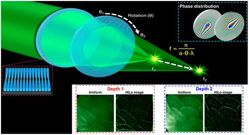
Fluorescence microscopy with optical sectioning capabilities is extensively utilized in biological research to obtain three-dimensional structural images of volumetric samples. Tunable lenses have been applied in microscopy for axial scanning to acquire multiplane images. However, images acquired by conventional tunable lenses suffer from spherical aberration and distortions. Here, we design, fabricate, and implement a dielectric Moiré metalens for fluorescence imaging. The Moiré metalens consists of two complementary phase metasurfaces, with variable focal length, ranging from ∼10 to ∼125 mm at 532 nm by tuning mutual angles. In addition, a telecentric configuration using the Moiré metalens is designed for high-contrast multiplane fluorescence imaging. The performance of our system is evaluated by optically sectioned images obtained from HiLo illumination of fluorescently labeled beads, as well as ex vivo mice intestine tissue samples. The compact design of the varifocal metalens may find important applications in fluorescence microscopy and endoscopy for clinical purposes.
中文翻译:

用于光学切片荧光显微镜的变焦元透镜
具有光学切片功能的荧光显微镜广泛用于生物学研究,以获得体积样品的三维结构图像。可调谐透镜已应用于显微镜中,用于轴向扫描以获取多平面图像。然而,由传统可调镜头获取的图像存在球面像差和畸变。在这里,我们设计、制造和实现了用于荧光成像的介电莫尔超透镜。莫尔超透镜由两个互补相位超表面组成,具有可变焦距,通过调整相互角度,在 532 nm 处的范围从 ~10 到 ~125 mm。此外,使用莫尔超透镜的远心配置专为高对比度多平面荧光成像而设计。我们系统的性能通过从荧光标记珠的 HiLo 照明以及离体小鼠肠道组织样本中获得的光学切片图像进行评估。变焦元透镜的紧凑设计可能会在荧光显微镜和内窥镜检查中找到重要的临床应用。
更新日期:2021-06-23
中文翻译:

用于光学切片荧光显微镜的变焦元透镜
具有光学切片功能的荧光显微镜广泛用于生物学研究,以获得体积样品的三维结构图像。可调谐透镜已应用于显微镜中,用于轴向扫描以获取多平面图像。然而,由传统可调镜头获取的图像存在球面像差和畸变。在这里,我们设计、制造和实现了用于荧光成像的介电莫尔超透镜。莫尔超透镜由两个互补相位超表面组成,具有可变焦距,通过调整相互角度,在 532 nm 处的范围从 ~10 到 ~125 mm。此外,使用莫尔超透镜的远心配置专为高对比度多平面荧光成像而设计。我们系统的性能通过从荧光标记珠的 HiLo 照明以及离体小鼠肠道组织样本中获得的光学切片图像进行评估。变焦元透镜的紧凑设计可能会在荧光显微镜和内窥镜检查中找到重要的临床应用。


 京公网安备 11010802027423号