Arthropod Structure & Development ( IF 1.7 ) Pub Date : 2021-05-08 , DOI: 10.1016/j.asd.2021.101055 Stefan Fischer 1 , Michael Laue 2 , Carsten H G Müller 3 , Ian A Meinertzhagen 4 , Hans Pohl 5

|
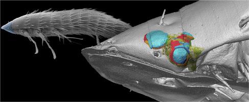
Stemmata of strepsipteran insects represent the smallest arthropod eyes known, having photoreceptors which form fused rhabdoms measuring an average size of 1.69 × 1.21 × 1.04 μm and each occupying a volume of only 0.97–1.16 μm3. The morphology of the stemmata of the extremely miniaturized first instar larva of Stylops ovinae (Strepsiptera, Stylopidae) was investigated using serial-sectioning transmission electron microscopy (ssTEM). Our 3D reconstruction revealed that, despite different proportions, all three stemmata maintain the same organization: a biconvex corneal lens, four corneagenous cells and five photoreceptor (retinula) cells. No pigment-containing cell-types were found to adjoin the corneagenous cells. Whereas the retinula cells are adapted to the limited space by having laterally bulged median regions, containing mitochondria and the smallest nuclei yet reported for arthropods (1.37 μm3), special adaptations are found in the corneagenous cells which have cell volumes down to 1 μm3. The corneagenous cells lack nuclei and pigment granules and bear only a few mitochondria (up to three) or none at all. Morphological adaptations due to miniaturization are discussed in the context of photoreceptor function and the visual needs of the larva.
中文翻译:

已知最小昆虫光感受器的超微结构 3D 重建:链翅目(六足目)一龄幼虫的茎
strepsipteran 昆虫的干细胞代表已知最小的节肢动物眼睛,具有形成平均尺寸为 1.69 × 1.21 × 1.04 μm 的融合横纹肌的感光器,每个仅占据 0.97–1.16 μm 3的体积。Stylops ovinae极小型化的一龄幼虫茎的形态(Streppsiptera, Stylopidae) 使用连续切片透射电子显微镜 (ssTEM) 进行研究。我们的 3D 重建显示,尽管比例不同,但所有三个干细胞都保持相同的组织:一个双凸角膜晶状体、四个角膜细胞和五个光感受器(视网膜)细胞。没有发现含有色素的细胞类型与角质细胞相邻。虽然视网膜细胞通过具有横向凸起的中间区域来适应有限的空间,其中包含线粒体和迄今为止报道的节肢动物最小的细胞核 (1.37 μm 3 ),但在细胞体积低至 1 μm 3的角质细胞中发现了特殊的适应性. 角质层细胞没有细胞核和色素颗粒,只有少数线粒体(最多三个)或根本没有。在光感受器功能和幼虫的视觉需求的背景下讨论了由于小型化引起的形态学适应。


 京公网安备 11010802027423号